. 2019 Dec 20;294(51):19795-19803.
doi: 10.1074/jbc.RA119.010730. Epub 2019 Nov 13.

Structural basis of binding of homodimers of the nuclear receptor NR4A2 to selective Nur-responsive DNA elements

Affiliations

Structural basis of binding of homodimers of the nuclear receptor NR4A2 to selective Nur-responsive DNA elements

Longying Jiang et al. J Biol Chem. .

Abstract

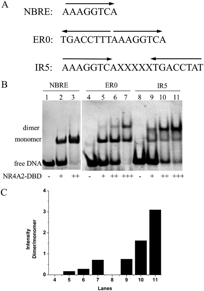
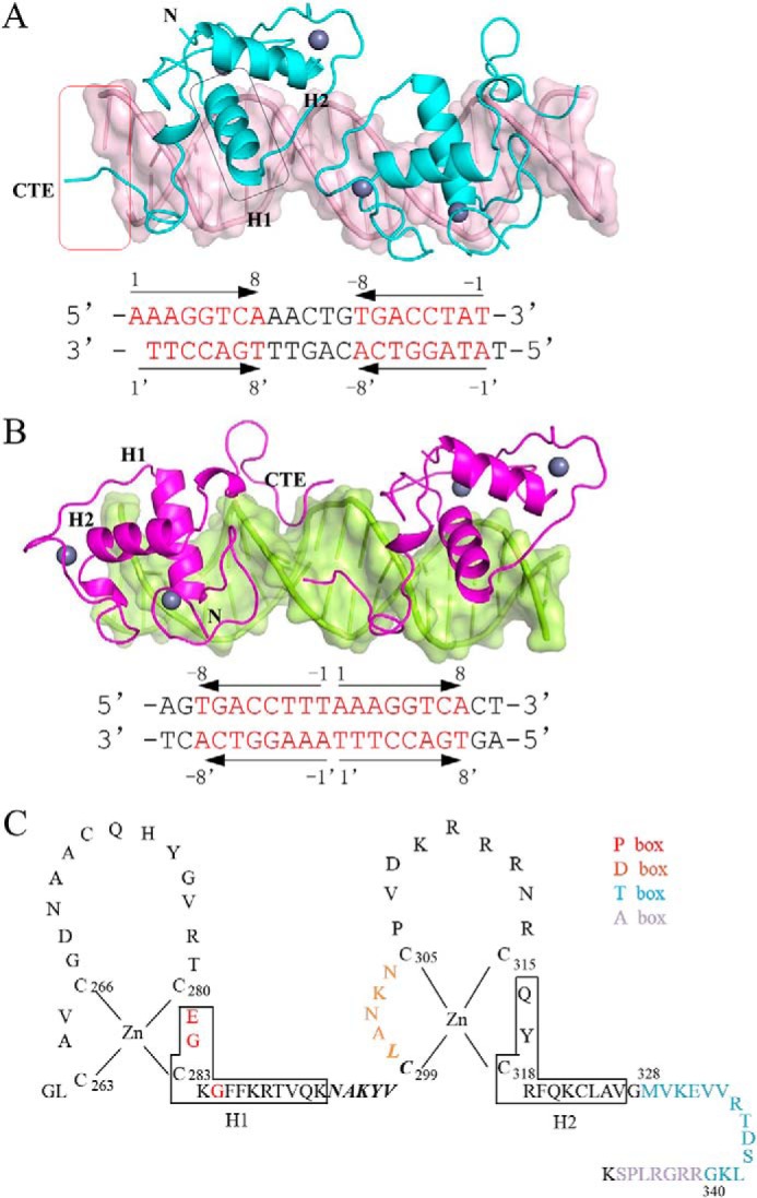
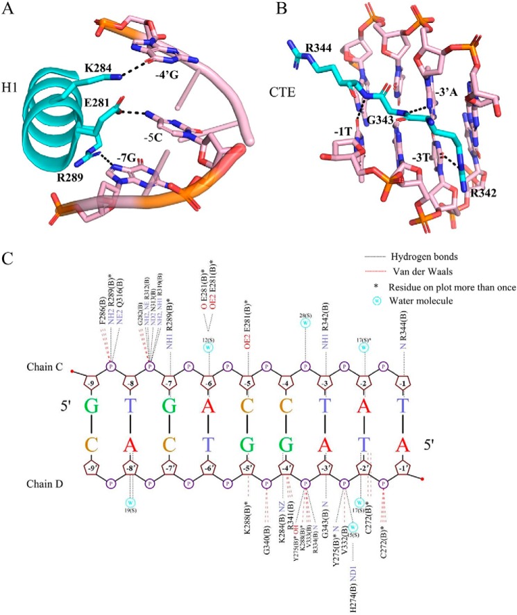
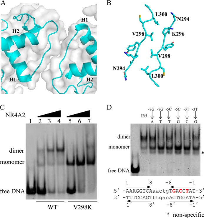
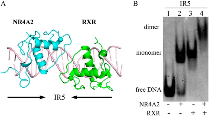
Proteins of nuclear receptor subfamily 4 group A (NR4A), including NR4A1/NGFI-B, NR4A2/Nurr1, and NR4A3/NOR-1, are nuclear transcription factors that play important roles in metabolism, apoptosis, and proliferation. NR4A proteins recognize DNA response elements as monomers or dimers to regulate the transcription of a variety of genes involved in multiple biological processes. In this study, we determined two crystal structures of the NR4A2 DNA-binding domain (NR4A2-DBD) bound to two Nur-responsive elements: an inverted repeat and an everted repeat at 2.6-2.8 Å resolution. The structures revealed that two NR4A2-DBD molecules bind independently to the everted repeat, whereas two other NR4A2-DBD molecules form a novel dimer interface on the inverted repeat. Moreover, substitution of the interfacial residue valine 298 to lysine as well as mutation of DNA bases involved in the interactions abolished the dimerization. Overall, our structural, biochemical, and bioinformatics analyses provide a molecular basis for the binding of the NR4A2 protein dimers to NurREs and advance our understanding of the dimerization specificity of nuclear receptors.

Keywords: Nur-responsive elements; crystal structure; dimerization; gene regulation; nuclear receptor; nuclear receptor subfamily 4 group A member 2; protein crystallization; protein–DNA interaction; protein–protein interaction; transcription factor.


Conflict of interest statement

The authors declare that they have no conflicts of interest with the contents of this article

Figures

Figure 1.
Figure 1.
Binding ability of NR4A2-DBD with different DNAs. A, DNA sequences of the response elements for NR4A2. B, binding features of NR4A2-DBD with different DNAs were determined by EMSA. Lanes 1, 4, and 8 show free DNA without protein. The primary concentrations of DNA and protein used were both 45 μm. + indicates that the molar ratio of protein to DNA is 1:1, and ++ indicates that the molar ratio is 2:1. C, quantification of the intensity of dimer/monomer bands. The graph shows the relative density of the dimer/monomer bands detected by EMSA.
Figure 2.
Figure 2.
Overall structures of NR4A2-DBD–DNA complexes. A, overall structure of the NR4A2-DBD–IR5 complex. NR4A2-DBD is colored cyan, and DNA targets are colored light pink. B, overall structure of the NR4A2-DBD–ER0 complex. NR4A2-DBD is colored magenta, and ER0 is colored lemon. The sequences of the DNA duplex used in the structures are listed below, and the core octanucleotides are colored red. The arrows show the orientation of each core half-site. Secondary structure elements (H1 and H2) are labeled, and the region in the box is involved in the protein–DNA interactions. C, amino acid sequences of NR4A2-DBD. Arrangement of residues 261–348 of NR4A2 shows the classic nuclear receptor-type zinc-finger motif and a CTE. The red and orange letters correspond to the P-box and D-box, respectively. The blue and purple letters correspond to the T-box and A-box, respectively. Solid-line boxes indicate α-helical segments. The residues in bold and italic are involved in dimerization.
Figure 3.
Figure 3.
Detailed protein–base contacts in the NR4A2-DBD–IR5 structure. A, stereo diagram of major groove recognition by helix H1 of NR4A2-DBD. B, stereo diagram of minor groove recognition by the C-terminal extension of NR4A2-DBD. The color is as shown in Fig. 2. All hydrogen bonds are shown as black dotted lines. C, summary of protein–DNA interactions between the NR4A2-DBD and the upstream half-site DNA generated by NUCPLOT. Hydrogen bonds are shown as black dotted lines. van der Waals interactions are shown as red dotted lines. Asterisks indicate residues that are on the plot more than once. W, water molecule.
Figure 4.
Figure 4.
Importance of protein–protein interactions and protein–DNA interactions in the NR4A2-DBD–IR5 structure. A, surface representation of the dimerization interface in two NR4A2-DBD molecules. B, detailed stereo diagram of the residues (sticks) involved in the dimer interface. C, the DNA binding ability of the WT and V298K variant of NR4A2-DBD was measured by EMSA. The concentration of the DNA and proteins used here was 45 μm. D, binding features of NR4A2-DBD with different IR5 DNA variants. The core sequences of the IR5 DNA duplex used are listed. The arrows show the orientation of each core half-site. The bases in red and bold are involved in the base–protein interaction and are mutated in the EMSA. The concentration of DNA used here were 45 μm, and the proteins were used at 90 μm.
Figure 5.
Figure 5.
Analysis of the NR4A2–RXR heterodimer. A, model of the NR4A2–RXR heterodimer on IR5. The RXR structure was obtained from a previously reported RXR–DR1 structure (PDB code 4CN2) and superimposed onto our NR4A2–IR5 complex structure. The RXR and NR4A2 structures were aligned to create the NR4A2–RXR–DNA structural model. C, DNA binding ability of the NR4A2–RXR heterodimer bound to DNAs.
Figure 6.
Figure 6.
Occurrence of the IR5 and ER0 binding motifs in NR4A-binding sites in human cells. A, analysis of the ratio of motifs IR5 and ER0 in the NR4A ChIP-seq data. B, representative gene promoters near NR4A-binding sites containing motif IR5.

References

    1. Wang J., Yang J., Zou Y., Huang G. L., and He Z. W. (2013) Orphan nuclear receptor nurr1 as a potential novel marker for progression in human prostate cancer. Asian Pac. J. Cancer Prev. 14, 2023–2028 10.7314/APJCP.2013.14.3.2023 - DOI - PubMed
    1. Mohan H. M., Aherne C. M., Rogers A. C., Baird A. W., Winter D. C., and Murphy E. P. (2012) Molecular pathways: the role of NR4A orphan nuclear receptors in cancer. Clin. Cancer Res. 18, 3223–3228 10.1158/1078-0432.CCR-11-2953 - DOI - PubMed
    1. Zhan Y. Y., Chen Y., Zhang Q., Zhuang J. J., Tian M., Chen H. Z., Zhang L. R., Zhang H. K., He J. P., Wang W. J., Wu R., Wang Y., Shi C., Yang K., Li A. Z., et al. (2012) The orphan nuclear receptor Nur77 regulates LKB1 localization and activates AMPK. Nat. Chem. Biol. 8, 897–904 10.1038/nchembio.1069 - DOI - PubMed
    1. Berwick D. C., and Harvey K. (2014) The regulation and deregulation of Wnt signaling by PARK genes in health and disease. J. Mol. Cell Biol. 6, 3–12 10.1093/jmcb/mjt037 - DOI - PMC - PubMed
    1. Yin K., Chhabra Y., Tropée R., Lim Y. C., Fane M., Dray E., Sturm R. A., and Smith A. G. (2017) NR4A2 promotes DNA double-strand break repair upon exposure to UVR. Mol. Cancer Res. 15, 1184–1196 10.1158/1541-7786.MCR-17-0002 - DOI - PubMed

Publication types

LinkOut - more resources