

Multiplex analysis of 40 cytokines do not allow separation between endometriosis patients and controls
- PMID: 31723213
- PMCID: PMC6853932
- DOI: 10.1038/s41598-019-52899-8
Multiplex analysis of 40 cytokines do not allow separation between endometriosis patients and controls
Abstract
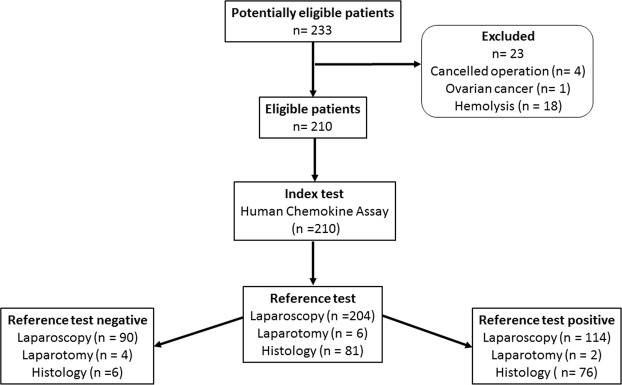
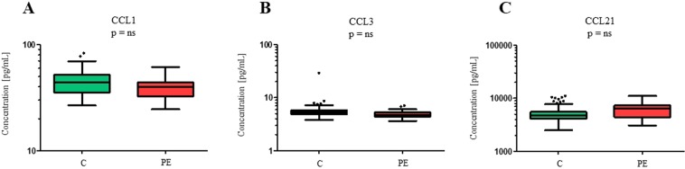
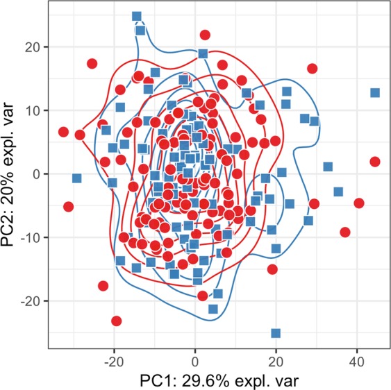
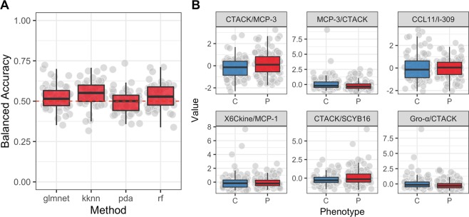
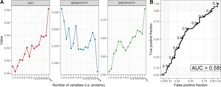
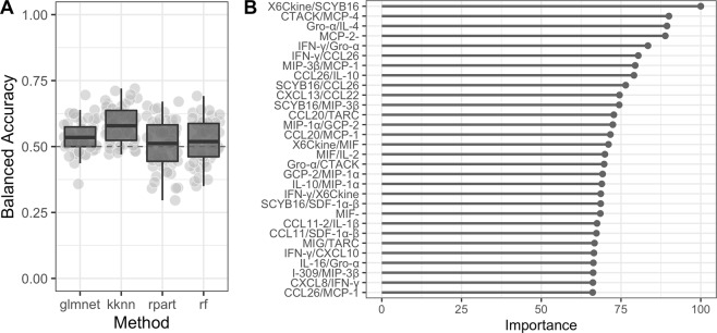
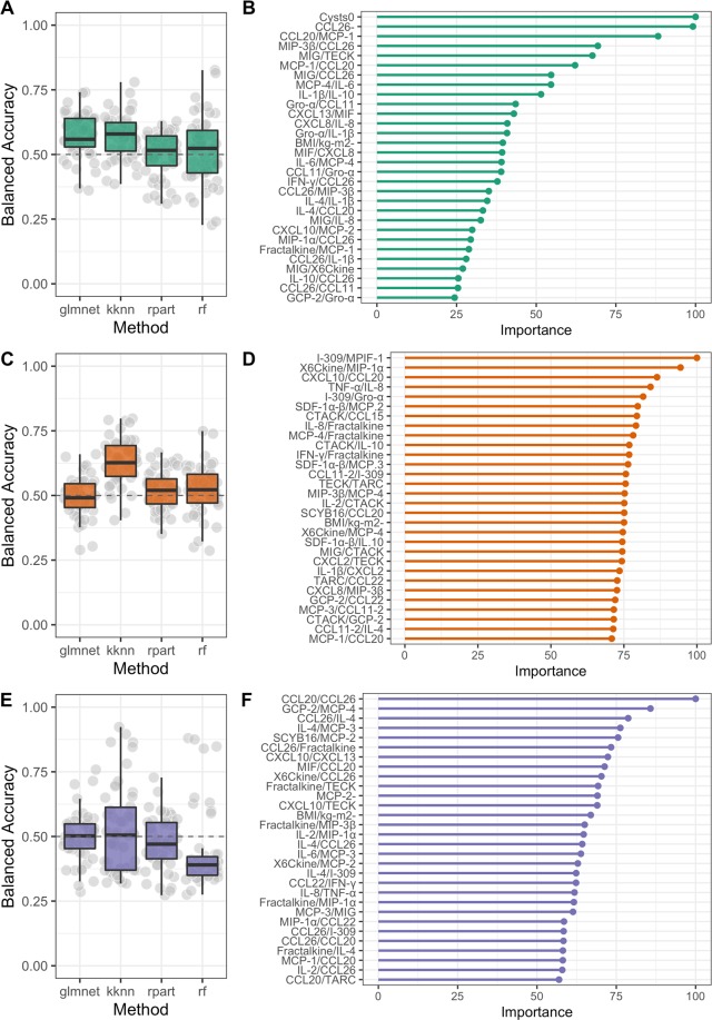
Endometriosis is a common gynaecological condition characterized by severe pelvic pain and/or infertility. The combination of nonspecific symptoms and invasive laparoscopic diagnostics have prompted researchers to evaluate potential biomarkers that would enable a non-invasive diagnosis of endometriosis. Endometriosis is an inflammatory disease thus different cytokines represent potential diagnostic biomarkers. As panels of biomarkers are expected to enable better separation between patients and controls we evaluated 40 different cytokines in plasma samples of 210 patients (116 patients with endometriosis; 94 controls) from two medical centres (Slovenian, Austrian). Results of the univariate statistical analysis showed no differences in concentrations of the measured cytokines between patients and controls, confirmed by principal component analysis showing no clear separation amongst these two groups. In order to validate the hypothesis of a more profound (non-linear) differentiating dependency between features, machine learning methods were used. We trained four common machine learning algorithms (decision tree, linear model, k-nearest neighbour, random forest) on data from plasma levels of proteins and patients' clinical data. The constructed models, however, did not separate patients with endometriosis from the controls with sufficient sensitivity and specificity. This study thus indicates that plasma levels of the selected cytokines have limited potential for diagnosis of endometriosis.
Conflict of interest statement
The authors declare no competing interests.
Figures
Similar articles
-
Identification of Serum Biomarkers for Diagnosis of Endometriosis Using Multiplex Immunoassays.Reprod Sci. 2020 May;27(5):1139-1147. doi: 10.1007/s43032-019-00124-2. Epub 2020 Jan 6. Reprod Sci. 2020. PMID: 32046464
-
Evaluation of a panel of 28 biomarkers for the non-invasive diagnosis of endometriosis.Hum Reprod. 2012 Sep;27(9):2698-711. doi: 10.1093/humrep/des234. Epub 2012 Jun 26. Hum Reprod. 2012. PMID: 22736326
-
Panels of cytokines and other secretory proteins as potential biomarkers of ovarian endometriosis.J Mol Diagn. 2015 May;17(3):325-34. doi: 10.1016/j.jmoldx.2015.01.006. Epub 2015 Mar 20. J Mol Diagn. 2015. PMID: 25797583
-
Biomarkers of endometriosis.Fertil Steril. 2013 Mar 15;99(4):1135-45. doi: 10.1016/j.fertnstert.2013.01.097. Epub 2013 Feb 13. Fertil Steril. 2013. PMID: 23414923 Review.
-
Biomarkers for the Noninvasive Diagnosis of Endometriosis: State of the Art and Future Perspectives.Int J Mol Sci. 2020 Mar 4;21(5):1750. doi: 10.3390/ijms21051750. Int J Mol Sci. 2020. PMID: 32143439 Free PMC article. Review.
Cited by
-
A first-in-class, non-invasive, immunodynamic biomarker approach for precision immuno-oncology.Oncoimmunology. 2022 Jan 10;11(1):2024692. doi: 10.1080/2162402X.2021.2024692. eCollection 2022. Oncoimmunology. 2022. PMID: 35036075 Free PMC article.
-
Evaluation of inflammatory serum parameters as a diagnostic tool in patients with endometriosis: a case-control study.Sci Rep. 2025 Jun 20;15(1):20172. doi: 10.1038/s41598-025-05719-1. Sci Rep. 2025. PMID: 40542028 Free PMC article.
-
Complement and coagulation cascade cross-talk in endometriosis and the potential of Janus Kinase inhibitors-a network meta-analysis.Front Immunol. 2025 Jul 8;16:1619434. doi: 10.3389/fimmu.2025.1619434. eCollection 2025. Front Immunol. 2025. PMID: 40698088 Free PMC article.
-
Proteomic analysis of peritoneal fluid identified COMP and TGFBI as new candidate biomarkers for endometriosis.Sci Rep. 2021 Oct 22;11(1):20870. doi: 10.1038/s41598-021-00299-2. Sci Rep. 2021. PMID: 34686725 Free PMC article.
-
Endometriosis in transgender men: recognizing the missing pieces.Front Med (Lausanne). 2023 Aug 31;10:1266131. doi: 10.3389/fmed.2023.1266131. eCollection 2023. Front Med (Lausanne). 2023. PMID: 37720510 Free PMC article.
References
-
- Revised American Society for Reproductive Medicine classification of endometriosis: 1996. Fertil. Steril. 67, 817–821 (1997). - PubMed
Publication types
MeSH terms
Substances
LinkOut - more resources
Full Text Sources
Medical
Research Materials
