

Dermoscopic-Histopathological Correlation of Eccrine Poroma: An Observational Study
- PMID: 31723462
- PMCID: PMC6830555
- DOI: 10.5826/dpc.0904a07
Dermoscopic-Histopathological Correlation of Eccrine Poroma: An Observational Study
Abstract
Background: Eccrine poroma (EP) is a benign adnexal neoplasm that can be pigmented in 17% of cases. Four histopathological variants of EP exist. Dermoscopically, EP can mimic many other skin neoplasms.
Objectives: To provide a dermoscopic-histopathological correlation of EP, classifying the clinical and dermoscopic features of EPs on the basis of their histopathological subtype, in an attempt to better characterize these entities.
Patients and methods: A single-center retrospective study was conducted. Clinical data were collected; patients were classified on the basis of the 4 histopathological variants of EPs. Dermoscopic images were reviewed. A dermoscopic-histopathological correlation was performed, and the results were compared with literature data.
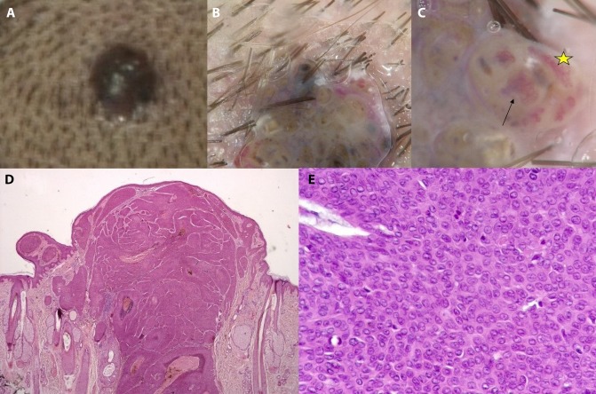
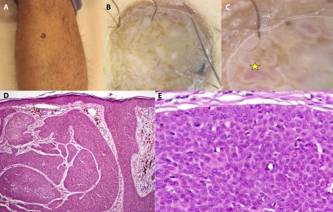
Results: Twenty-six lesions were included, both pigmented and nonpigmented. Three of the 4 histopathological variants were identified. Different dermoscopic features were observed for each distinct histopathological subtype of EP. The lesions mimicked different types of other skin neoplasms, in particular: nonpigmented hidroacanthoma simplex resembled nonmelanoma skin cancer; pigmented hidroacanthoma simplex appeared like a seborrheic keratosis or a solar lentigo; EPs sensu stricto presented as pink nodules if nonpigmented and were similar to seborrheic keratosis if pigmented; dermal duct tumors appeared as pigmented nodular lesions.
Conclusions: Distinct dermoscopic features appeared to be recurrent in each histopathological variant. Dermoscopy can provide important clues for the diagnosis of EP; the final diagnosis is allowed by histopathology. To achieve a correct diagnosis of EP, because of its clinical and dermoscopic variability, surgical excision is recommended.
Keywords: dermoscopy; diagnosis; eccrine; histopathology; poroma.
Copyright: ©2019 Chessa et al.
Conflict of interest statement
Competing interests: The authors have no conflicts of interest to disclose.
Figures


References
-
- Minagawa A, Koga H. Dermoscopy of pigmented poromas. Dermatology. 2010;221(1):78–83. - PubMed
-
- Lallas A, Chellini PR, Guimarães MG, et al. Eccrine poroma: the great dermoscopic imitator. J Eur Acad Dermatol Venereol. 2016;30(10):e61–e63. - PubMed
-
- Battistella M, Langbein L, Peltre B, Cribier B. From hidroacanthoma simplex to poroid hidradenoma: clinicopathologic and immunohistochemic study of poroid neoplasms and reappraisal of their histogenesis. Am J Dermatopathol. 2010;32(5):459–468. - PubMed
-
- Martín JM, Bella-Navarro R, Jordá E. Vascular patterns in dermoscopy. Actas Dermosifiliogr. 2012;103(5):357–375. - PubMed
LinkOut - more resources
Full Text Sources