. 2019 Aug 12;5(8):3935-3943.
doi: 10.1021/acsbiomaterials.8b01359. Epub 2019 Feb 6.

Directed Collective Cell Migration Using Three-Dimensional Bioprinted Micropatterns on Thermoresponsive Surfaces for Myotube Formation

Affiliations

Directed Collective Cell Migration Using Three-Dimensional Bioprinted Micropatterns on Thermoresponsive Surfaces for Myotube Formation

Wenqiang Du et al. ACS Biomater Sci Eng. .

Abstract

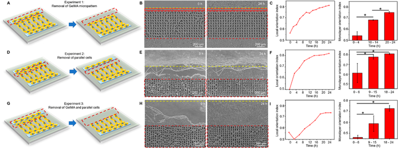
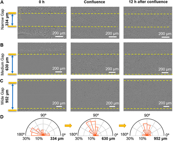
Directed collective cell migration governs cell orientation during tissue morphogenesis, wound healing, and tumor metastasis. Unfortunately, current methods for initiating collective cell migration, such as scratching, laser ablation, and stencils, either introduce uncontrolled cell-injury, involve multiple fabrication processes, or have utility limited to cells with strong cell-cell junctions. Using three-dimensional (3D) bioprinted gelatin methacryloyl (GelMA) micropatterns on temperature-responsive poly(N-isopropylacrylamide) (PNIPAm) coated interfaces, we demonstrate that directed injury-free collective cell migration could occur in parallel and perpendicular directions. After seeding cells, we created cell-free spaces between two 3D bioprinted GelMA micropatterns by lowering the temperature of PNIPAm interfaces to promote the cell detachment. Unlike conventional collective cell migration methods initiated by stencils, we observed well-organized cell migration in parallel and perpendicular to 3D bioprinted micropatterns as a function of the distance between 3D bioprinted micropatterns. We further established the utility of controlled collective cell migration for directed functional myotube formation using 3D bioprinted fingerprintlike micropatterns as well as iris musclelike concentric circular patterns. Our platform is unique for myoblast alignment and myotube formation because it does not require anisotropic guidance cues. Together, our findings establish how to achieve controlled collective cell migration, even at the macroscale, for tissue engineering and regeneration.

Keywords: 3D printing; directed collective cell migration; myotube orientation; temperature-responsive surface.


Figures

Figure 1.
Figure 1.
3D bioprinted hydrogel micropatterns to promote directed collective cell migration. (A) Schema (top) and a picture (bottom) of 3D bioprinted GelMA micropatterns on PNIPAm coated surface. (B) Uniform cell seeding on GelMA micropatterns and PNIPAm coated surface at 37 °C. (C) Detachment of cells to generate cell-free space by reducing temperature of thermoresponsive PNIPAm coated interface to 20 °C. (D) Initiation of directed collective cell migration by 3D bioprinted micropatterns at 37 °C. Yellow lines with arrowheads show cell migration directions parallel or perpendicular to 3D bioprinted micropatterns. (E) Directed collective myotube fibers’ orientation after changing the growth media to differentiation media. Yellow lines with arrowheads show myotube orientation directions either parallel or perpendicular to 3D bioprinted micropatterns.
Figure 2.
Figure 2.
Directed collective cell migration in parallel and perpendicular directions initiated by a line micropattern as a function of distance from the boundary. (A) Time-lapse images of directed collective cell migration in 72 h. The micropattern boundary is indicated as a yellow dotted line. Cell migration perpendicular (red arrowhead) and parallel (blue line with double arrowheads) to the micropattern boundary is indicated at a different distance (red dotted line) from the GelMA boundary over time. The perpendicular direction to the micropattern boundary is defined as 0°. (B–C) Orientation maps and orientation index of collectively migrating cells in the perpendicular direction over time and at different distances. Cells located along the micropattern boundary are not calculated. (D) Orientation index of cells migrating away, in the perpendicular direction, from the micropattern boundary at different distances, suggesting cell migration direction transition from parallel to perpendicular after 200 μm which becomes perpendicular after 400 μm from the boundary. Thus, cell migration is a function of the distance from the boundary of the GelMA micropattern. (E) Orientation maps and orientation index of collectively migrating cells, located along the micropattern boundary, in the parallel direction over time, suggesting that cell migration is not a function of time. The wind rose maps show the overlapped orientation angle distribution. (F) Velocity cloud map of collective migration of cells in the perpendicular direction after reaching confluence from 72 to 80 h. (G) Mean velocity orientation map of (F). (H–I) Velocity magnitude and orientation as a function of distance from the micropattern boundary. *p < 0.05.
Figure 3.
Figure 3.
Parallel collective cell migration requires the presence of micropattern whereas perpendicular collective cell migration is preserved after initiation. (A) Schema demonstrating removal of the GelMA micropattern. (B) Time-lapse images of collective migration after removal of the GelMA micropattern. Red dotted rectangles indicate the region for calculating local orientation index. The red rectangle indicates the parallel cells. Yellow dotted lines indicate the boundary of micropatterns. (C) Local orientation index as a function of culture time after removal of GelMA (left). Overall orientation index of cell monolayer as a function of time after removal of GelMA (right). (D) Schema demonstrating removal of a lining of parallel cells adjacent to the GelMA micropattern (and not GelMA micropattern). (E) Time-lapse images of collective migration after removal of a lining of parallel cells adjacent to the GelMA micropattern. (F) Local orientation index as a function of culture time after removal of a lining of parallel cells adjacent to the GelMA micropattern (left). Overall orientation index of cell monolayer as a function of time after removal of a lining of parallel cells adjacent to the GelMA micropattern (right). (G) Schema demonstrating removal of both the GelMA micropattern and alining of parallel cells adjacent to the GelMA micropattern. (H) Time-lapse images of collective migration after removal of both the GelMA micropattern and a lining of parallel cells adjacent to the GelMA micropattern. (I) Local orientation index as a function of culture time after removal of both the GelMA micropattern and a lining of parallel cells adjacent to the GelMA micropattern (left). Monolayer orientation index of cell monolayer as a function of time after removal of both the GelMA micropattern and a lining of parallel cells adjacent to the GelMA micropattern (right). Each experiment was repeated at least three times. *p < 0.05.
Figure 4.
Figure 4.
Directed collective cell migration as a function of a distance between two GelMA micropatterns. (A–C) Time-lapse images of directed collective cell migration initiated by two micropatterns separated by narrow (334 μm), medium (630 μm), and wide (952 μm) distances. (D) Cell orientation distribution for micropatterns separated by narrow (334 μm), medium (630 μm), and wide (952 μm) gaps at 12 h after confluence.
Figure 5.
Figure 5.
Directed collective cell migration of functional myotubes in perpendicular and parallel directions at millimeter-scale. (A) Schema of directed myoblast migration followed by myotube formation. (B–C) Immunofluorescent images show myosin (MHC, red) and nuclear (DAPI, light blue) staining in unpatterned and patterned myotube aligned after directed collective cell migration. Arrowheads indicate the position of the parallel myotubes after differentiation. Inserted image shows the sarcomere structure (green) in the myotube. (D–E) Myotube and nuclear orientation as well as fusion index of myotubes emerged from unpatterned (blue), parallel (green), and perpendicular (orange) collective cell migration. (F) Myotube contraction using 1–4 Hz electricpulse stimulation. (G) Fourier transform analysis ofthe myotube contraction frequency. (H) High resolution image showing N-cadherin (green) and DAPI (light blue) staining of myotubes. *p < 0.05.
Figure 6.
Figure 6.
Directed collective cell migration to establish a 3D bioprinted fingerprint with precise myotube orientation. (A–B) 3D model and G-code for fingerprint. (C) Immunofluorescent images of myotube (MHC), nuclei (DAPI), and merged in 3D bioprinted fingerprint pattern with different micropattern spacing. Blue arrows indicate the orientation of myotubes. Yellow lines indicate GelMA micropattern boundaries. (D–E) Myotube orientation angle and fusion index to demonstrate that the cell migration pattern is dependent on the distance between the GelMA micropattern. * p < 0.05.
Figure 7.
Figure 7.
3D bioprinted directed collective cell migration to engineer iris radial and circumferential muscle fibers. (A) Human iris radial muscle fiber (top) was reverse-engineered in an iris-mimic structure (bottom). (B) G-code for 3D printing of an iris. (C) Schema shows relative orientation of radial and circular muscle fibers in the human iris. (D) Engineering of patterned radial and circumferential muscle fibers to mimic human iris (left). Zoomed in (2×) image shows the interface of radial and circumferential muscle fibers (right). Yellow dotted lines indicate micropattern boundaries. White and blue arrows indicate the radial and circumferential muscle fibers. (E) Zoomed in (3×) image of muscle fiber orientation in human iris as seen in A. (F) Distribution of the orientation angle of radial muscle fibers in engineered 3D bioprinted iris (red) and in human iris (blue).

References

    1. Haeger A; Wolf K; Zegers MM; Friedl P Collective cell migration: guidance principles and hierarchies. Trends Cell Biol. 2015, 25 (9), 556–566. - PubMed
    1. Montell DJ; Yoon WH; Starz-Gaiano M Group choreography: mechanisms orchestrating the collective movement of border cells. Nat. Rev. Mol. Cell Biol. 2012, 13, 631. - PMC - PubMed
    1. Vedula SRK; Hirata H; Nai MH; Brugues A; Toyama Y; Trepat X; Lim CT; Ladoux B Epithelial bridges maintain tissue integrity during collective cell migration. Nat. Mater. 2014,13 (1), 87–96. - PubMed
    1. Scarpa E; Mayor R Collective cell migration in development. J. Cell Biol. 2016, 212 (2), 143–155. - PMC - PubMed
    1. Londono C; Loureiro MJ; Slater B; Lucker PB; Soleas J; Sathananthan S; Aitchison JS; Kabla AJ; McGuigan AP Nonautonomous contact guidance signaling during collective cell migration. Proc. Natl. Acad. Sci. U.S.A. 2014, 111 (5), 1807–1812. - PMC - PubMed

LinkOut - more resources