

Extensive and Progressive Cerebral Infarction after Mycoplasma pneumoniae Infection
- PMID: 31723636
- PMCID: PMC6786714
- DOI: 10.4266/kjccm.2016.00283
Extensive and Progressive Cerebral Infarction after Mycoplasma pneumoniae Infection
Abstract
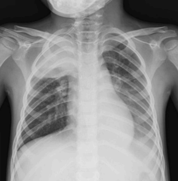
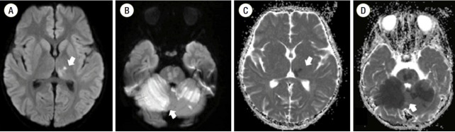
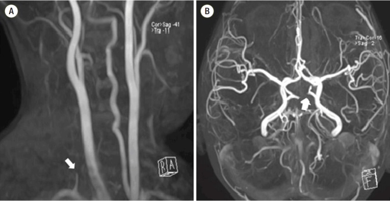
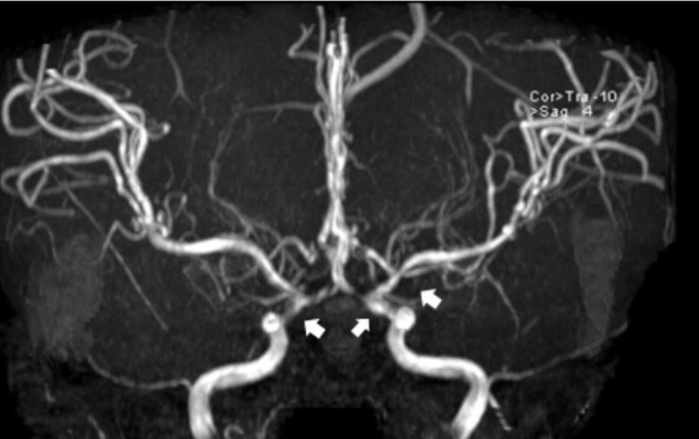
Acute cerebral infarctions are rare in children, however they can occur as a complication of a Mycoplasma pneumoniae (MP) infection due to direct invasion, vasculitis, or a hypercoagulable state. We report on the case of a 5-year-old boy who had an extensive stroke in multiple cerebrovascular territories 10 days after the diagnosis of MP infection. Based on the suspicion that the cerebral infarction was associated with a macrolide-resistant MP infection, the patient was treated with levofloxacin, methyl-prednisolone, intravenous immunoglobulin, and enoxaparin. Despite this medical management, cerebral vascular narrowing progressed and a decompressive craniectomy became necessary for the patient's survival. According to laboratory tests, brain magnetic resonance imaging, and clinical manifestations, the cerebral infarction in this case appeared to be due to the combined effects of hypercoagulability and cytokine-induced vascular inflammation.
Keywords: cerebral infarction; child; mycoplasma pneumoniae; thrombosis; vasculitis.
Copyright © 2017 The Korean Society of Critical Care Medicine.
Conflict of interest statement
No potential conflict of interest relevant to this article was reported.
Figures
References
-
- Tsiodras S, Kelesidis I, Kelesidis T, Stamboulis E, Giamarellou H. Central nervous system manifestations of Mycoplasma pneumoniae infections. J Infect. 2005;51:343–54. - PubMed
-
- Ovetchkine P, Brugieres P, Seradj A, Reinert P, Cohen R. An 8-old boy with acute stroke and radiological signs of cerebral vasculitis after recent Mycoplasma pneumoniae infection. Scand J Infect Dis. 2002;34:307–9. - PubMed
-
- Ryu JS, Kim HJ, Sung IY, Ko TS. Posterior cerebral artery occlusion after Mycoplasma pneumoniae infection associated with genetic defect of MTHFR C677T. J Child Neurol. 2009;24:891–4. - PubMed
-
- Leonardi S, Pavone P, Rotolo N, La Rosa M. Stroke in two children with Mycoplasma pneumoniae infection. A causal or casual relationship? Pediatr Infect Dis J. 2005;24:843–5. - PubMed
-
- Lee CY, Huang YY, Huang FL, Liu FC, Chen PY. Mycoplasma pneumoniae-associated cerebral infarction in a child. J Trop Pediatr. 2009;55:272–5. - PubMed
Publication types
LinkOut - more resources
Full Text Sources
