Multicenter Study
. 2020 Apr;44(4):467-476.
doi: 10.1097/PAS.0000000000001405.

Non-neoplastic Polyps of the Gallbladder: A Clinicopathologic Analysis of 447 Cases

Affiliations
Multicenter Study

Non-neoplastic Polyps of the Gallbladder: A Clinicopathologic Analysis of 447 Cases

Orhun C Taskin et al. Am J Surg Pathol. 2020 Apr.

Abstract

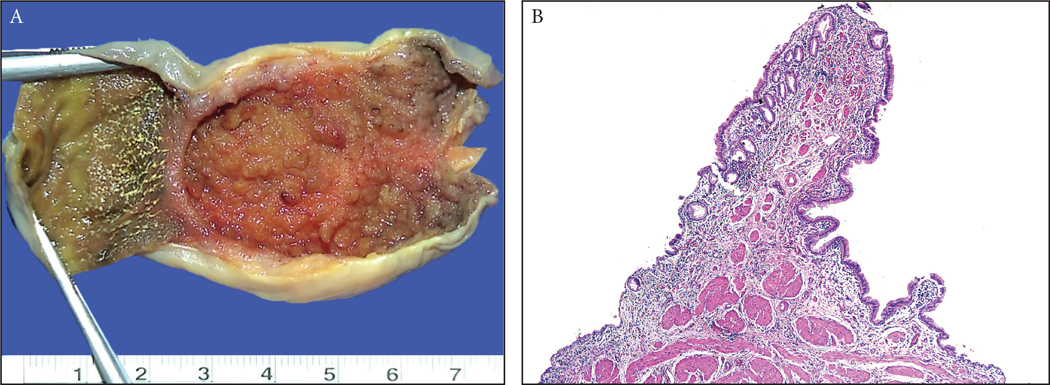
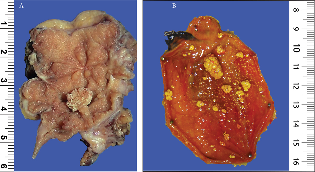
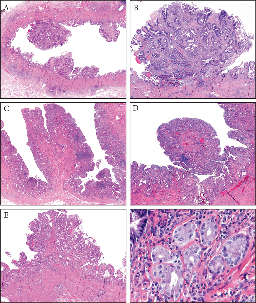
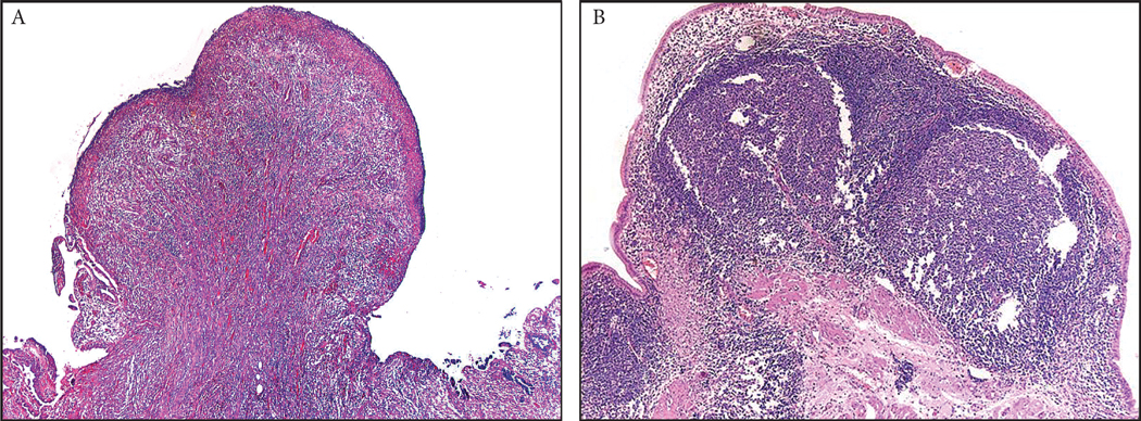
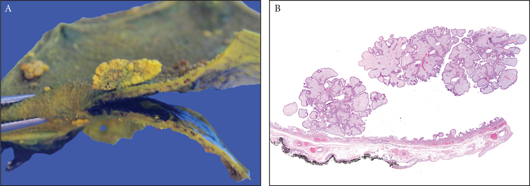
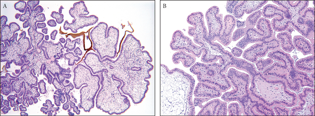
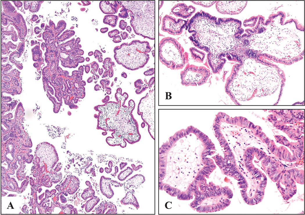
There is no systematic histopathologic analysis of non-neoplastic polyps in the gallbladder. In this study, in addition to a computer search for cases designated as "polyp," a systematic review of 2533 consecutive routinely sampled archival and 203 totally submitted prospective cholecystectomies were analyzed for >2 mm polyps (cut-off was based on radiologic sensitivity). A total of 447 non-neoplastic polyps were identified. The frequency was 3% in archival cases and 5% in totally submitted cases. Only 21 (5%) were ≥1 cm. The average age was 52 years, and the female to male ratio was 3.1. Two distinct categories were delineated: (1) injury-related polyps (n=273): (a) Fibro(myo)glandular polyps (n=214) were small (mean=0.4 cm), broad-based, often multiple (45%), almost always (98%) gallstone-associated, and were composed of a mixture of (myo)fibroblastic tissue/lobular glandular units with chronic cholecystitis. Dysplasia seen in 9% seemed to be secondary involvement. (b) Metaplastic pyloric glands forming polypoid collections (n=42). (c) Inflammatory-type polyps associated with acute/subacute injury (11 granulation tissue, 3 xanthogranulomatous, 3 lymphoid). (2) Cholesterol polyps (n=174) occurred in uninjured gallbladders, revealing a very thin stalk, edematous cores devoid of glands but with cholesterol-laden macrophages in 85%, and cholesterolosis in the uninvolved mucosa in 60%. Focal low-grade dysplasia was seen in 3%, always confined to the polyp, unaccompanied by carcinoma. In conclusion, non-neoplastic polyps are seen in 3% of cholecystectomies and are often small. Injury-related fibromyoglandular polyps are the most common. Cholesterol polyps have distinctive cauliflower architecture, often in a background of uninjured gallbladders with cholesterolosis and may lack the cholesterol-laden macrophages in the polyp itself. Although dysplastic changes can involve non-neoplastic polyps, they do not seem to be the cause of invasive carcinoma by themselves.


Conflict of interest statement

- Conflict of Interest: Authors declare that they have no conflict of interest to disclose.

Figures

Figure 1:
Figure 1:
(A) There are anatomic polypoid folds such as Phrigian cap deformity as well as the valves at the neck/cystic-duct region and those occur due to impacted stones, all of which were excluded from the study. Typically, these form a pencil-like projection that come out of the surface with an angle. (B) Microscopically, these tend to have a well defined and organized muscle core at its center that tapers towards the tip, and a normal mucosal covering that is almost equidistant from the muscle similar to the mucosa elsewhere.
Figure 2:
Figure 2:
(A) A 0.7 cm fibromyoglandular polyp is seen in the background of a polypoid/nodular gallbladder mucosa with prominent wall thickening and gallstones (See Figure 3 for microscopy). (B) A case of multiple cholesterol polyps, which were typically seen in gallbladders devoid of any significant chronic changes (notice the non-polypoid mucosa). Because of the presence of cholesterol-laden macrophages, which were often in abundance, they had the distinctive yellow color in macroscopic examination. They were characterized by a striking cauliflower pattern which was highly consistent in virtually all examples.
Figure 3:
Figure 3:
Fibromyoglandular polyps. (A) These occurred in the background of mucosal injury and were commonly multifocal. They had a spectrum of glandular and stromal components with variable muscle participation. The glands were also distributed variably. (B) Some had more fibrotic stroma and relatively evenly distriuted glands. (C) Others had substantial amount of muscular stroma with mucosally-oriented glandular components. (D) Inflammation can be prominent in some. (E) Lobulated collections of glands could be seen and (F) these were often of pyloric gland type. [Microscopic photographs of Figure 2a]
Figure 4:
Figure 4:
Inflammatory polyps constituted about 4% of all non-neoplastic polyps of the gallbladder. They were composed of various types of inflammatory process, including (A) granulation tissue, (B) lymphoid aggregates or xantogranulomas.
Figure 5:
Figure 5:
Cholesterol polyps were typically found in gallbladders that were devoid of any significant chronic changes. (A) They could be multifocal as seen in this example, though not as frequently as the fibromyoglandular polpys. (B) They had very thin stalks and as a result they commonly were detached from the surface. The main characteristic of cholesterol polyps, even more so than the cholesterol macrophages (lacking in 15% and barely visible in many others), was their distinctive cauliflower architecture. Another consistent finding was the conspicuous lack of epithelial elements in the cores of the polyp.
Figure 6:
Figure 6:
Cholesterol polyps were characterized by the cauliflower architecture, (A) even when the polyp acquired more complex architecture and (B) was devoid of lipid-laden macrophages.
Figure 7:
Figure 7:
Dysplastic type changes in a cholesterol polyp. This polyp shows the distinctive cauliflower pattern of a cholesterol polyp. The epithelium shows atypia that is substantially different than the normal epithelium that typically covers a cholesterol polyp. No substantial injury changes are noted to attribute the atypical changes to. This was classified as dysplasia in the original report, and was also classified as low-grade dysplasia also in this study although the precise nature of this process remains to be determined. No high-grade dysplasia or carcinoma is seen (and it was not seen in any of the other cases either).

References

    1. Wiles R, Thoeni RF, Barbu ST, et al. Management and follow-up of gallbladder polyps : Joint guidelines between the European Society of Gastrointestinal and Abdominal Radiology (ESGAR), European Association for Endoscopic Surgery and other Interventional Techniques (EAES), International Society of Digestive Surgery - European Federation (EFISDS) and European Society of Gastrointestinal Endoscopy (ESGE). Eur. Radiol. 2017;27:3856–3866. doi:10.1007/s00330-017-4742-y. - DOI - PMC - PubMed
    1. McCain RS, Diamond A, Jones C, et al. Current practices and future prospects for the management of gallbladder polyps: A topical review. World J. Gastroenterol. 2018;24:2844–2852. doi:10.3748/wjg.v24.i26.2844. - DOI - PMC - PubMed
    1. Babu BI, Dennison AR, Garcea G. Management and diagnosis of gallbladder polyps: a systematic review. Langenbeck’s Arch. Surg. 2015;400:455–62. doi:10.1007/s00423-015-1302-2. - DOI - PubMed
    1. Elmasry M, Lindop D, Dunne DFJ, et al. The risk of malignancy in ultrasound detected gallbladder polyps: A systematic review. Int. J. Surg. 2016;33 Pt A:28–35. doi:10.1016/j.ijsu.2016.07.061. - DOI - PubMed
    1. Albores-Saavedra J, Henson DE. Tumors of the Gallbladder and Extrahepatic Bile Ducts. In: Atlas of Tumor Pathology. 2nd series. Washington, DC: Armed Forces Institute of Pathology; 1986.

Publication types

MeSH terms