

Review
doi: 10.1021/acs.analchem.9b05080.
Epub 2019 Dec 2.
Multiplexed Immunosensors and Immunoarrays
Affiliations
- PMID: 31726821
- PMCID: PMC7202053
- DOI: 10.1021/acs.analchem.9b05080
Item in Clipboard
Review
Multiplexed Immunosensors and Immunoarrays
Anal Chem.
.
No abstract available
Conflict of interest statement
The authors declare no competing financial interest.
Figures
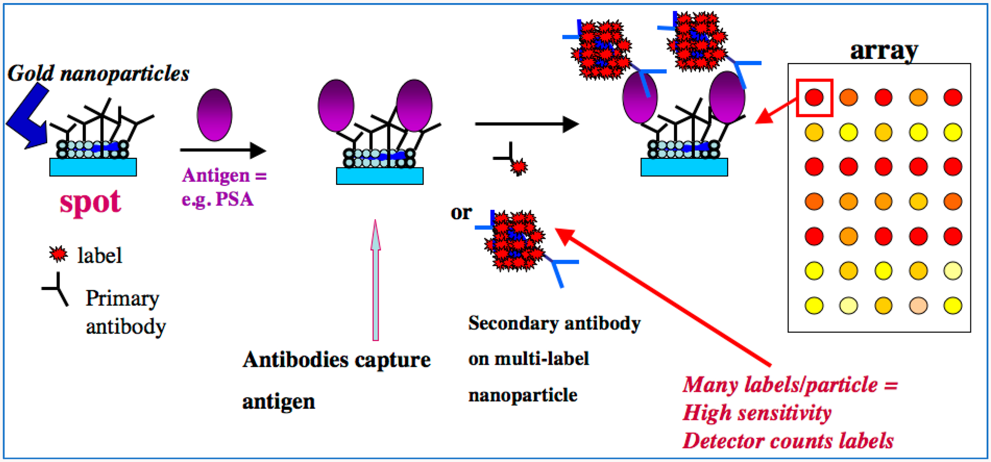
Example of a modern approach to sandwich immunoarrays. On left, a gold nanoparticle-decorated spot (to achieve high surface area) on the array is represented with attached primary antibodies (Ab1). Sample is delivered to the array, which can have a number of different Ab1 spots to capture a range of different antigens in a multiplexed assay. For our example SPOT, the antigen is prostate specific antigen, a biomarker protein for prostate cancer. The antigens are captured by Ab1’s on their specific spots, usually during an incubation period. After washing, secondary or detection antibodies (Ab2) are introduced, shown here by two examples. The conventional approach employs a single labeled antibody, while a more sensitive assay can be designed with multiple labels to amplify the signals. This step is followed by another incubation period, washing, and detection. These kinds of arrays can be integrated with microfluidics for sample and reagent delivery and automation.
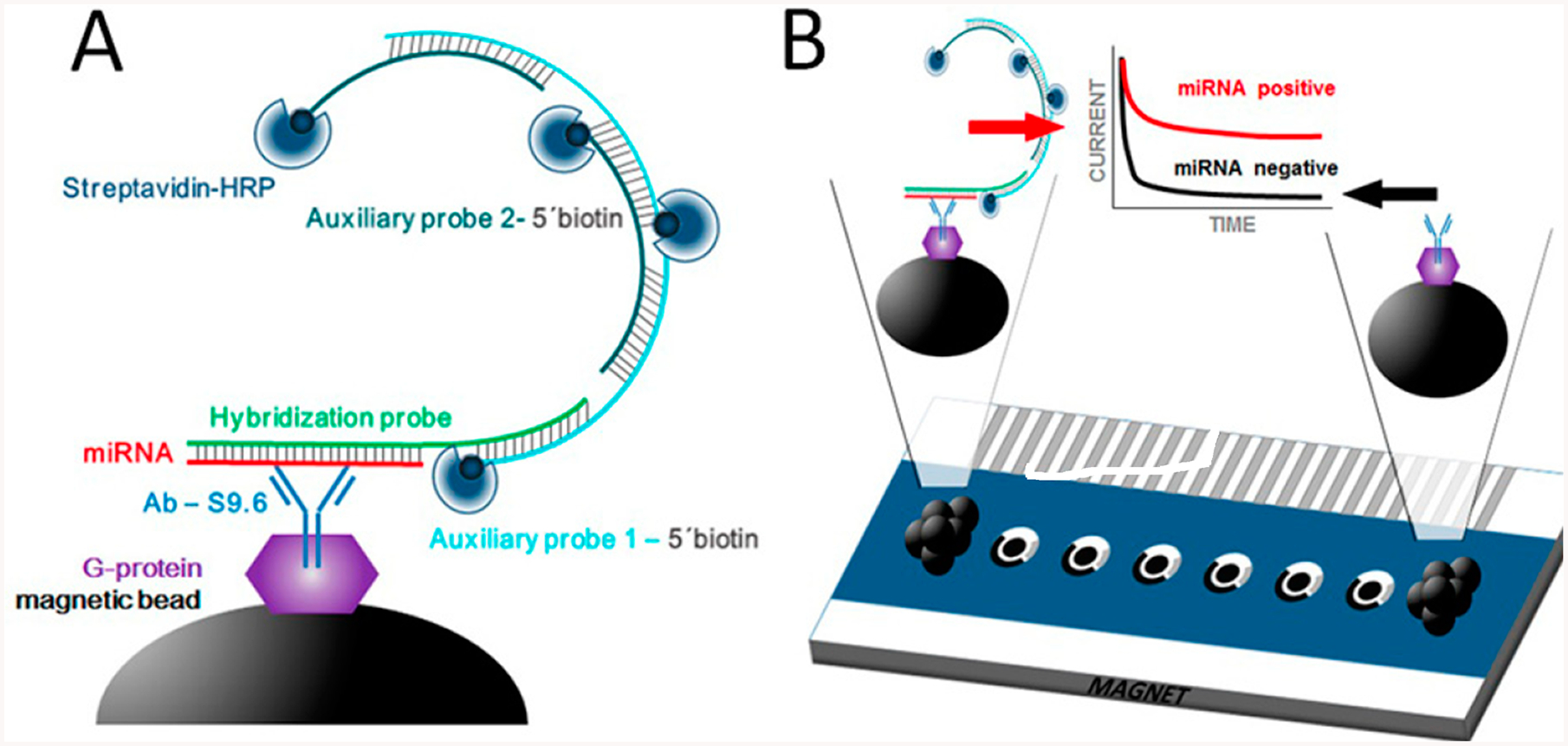
Schematic display of HCR-based miRNA immunosensing: (A) preparation of MBs including modification with AbS9.6, selective capture of the target miRNA/HP heteroduplex, HCR at the MBs by hybridization of the HP with AP1 and AP2 probes, and labeling of the biotin moieties with Strep-HRP. (B) Magnetic attraction of the modified MBs to the surface of the working electrodes (via magnet placed below the array) and chronoamperometric measurement of the enzymatic reduction of H2O mediated by hydroquinone. The cathodic current increased with the concentration of the target miRNA. Reproduced from Multiplexed Immunosensing Platform Coupled to Hybridization Chain Reaction for Electrochemical Determination of Micro RNAs in Clinical Samples, Jirakova, L.; Hrstka, R.; Campuzano, S.; Pingarrón, J.M.; Bartosik, M. Electroanalysis, Vol. 31, Issue 2 (ref 21). Copyright 2019 Wiley.
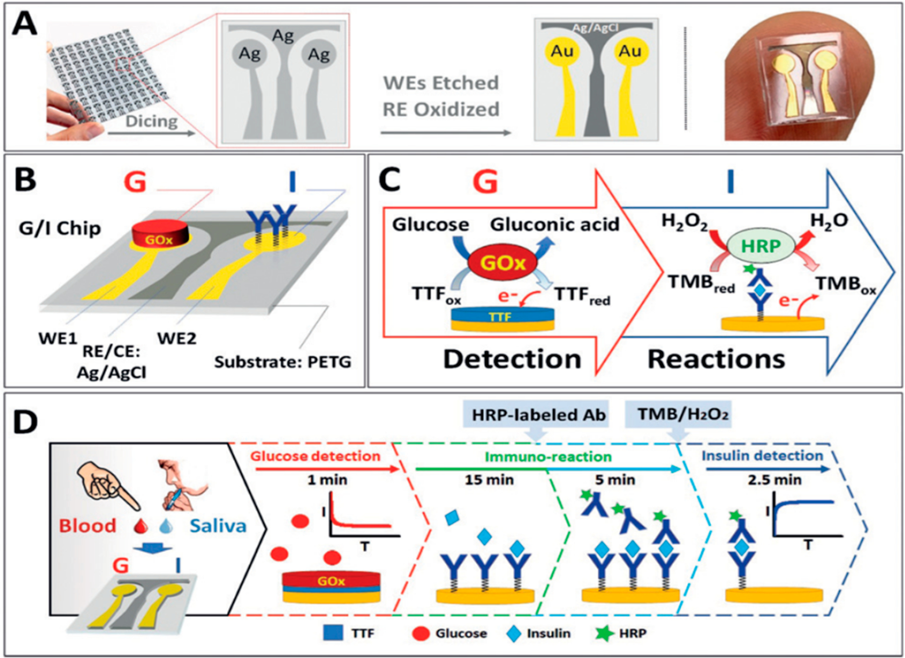
Dual G/I biosensor chip. (A) The G/I sensor chip array showing the two Au WEs for glucose and insulin biosensors, with Ag/AgCl electrode reference. (B) Localized detection of G and I on a sensor chip, showing the immobilized GOx and insulin antibody bioreceptors. (C) Recognition and redox processes in G/I sensing. Glucose detection is amperometric +0.2 V on AuWE1 with TTF-mediated biocatalytic (GOx) oxidation of G. Insulin is detected on Au WE2 at @0.1 V by sandwich immunoreaction assay by measuring H2O2 reduction current catalyzed HRP and mediated by 3,3′,5,5′-tetramethylbenzidine (TMB). (D) Glucose is measured followed by 15 min sample incubation and subsequent 5 min incubation with HRP-labeled antibody. Finally, insulin is monitored after washing the chip and adding mediator and H2O2. Reproduced from Enzymatic/Immunoassay Dual-Biomarker Sensing Chip: Toward Decentralized Insulin/Glucose Detection, Vargas, E.; Teymourian, H.; Tehrani, F.; Eksin, E.; Sánchez-Tirado, E.; Warren, P.; Erdem, A.; Dassau, E.; Wang, J. Angew. Chem. Int. Ed., Vol. 58, Issue 19 (ref 28). Copyright 2019 Wiley.
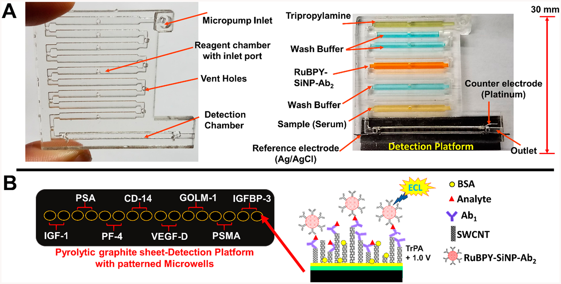
Representations of 3D-printed immunoarray: (A) 3D printed microfluidic array with chambers to hold sample, wash buffers, detection nanoparticles, and coreactant for ECL generation. Array is shown on the left without detection chip, and on the right bonded to a pyrolytic graphite sheet (PGS) microwell detection chip with reagent and sample chambers filled with dye solutions for visualization. (B) Representative disposable PGS chip with heat transferred microwells printed using hydrophobic toner ink. Inset illustrates a sandwich immunoassay on a single wall carbon nanotube forest (SWCNT) in one microwell. Reproduced from Kadimisetty, K.; Malla, S.; Bhalerao, K. S.; Mosa, I. M.; Bhakta, S.; Lee, N. H.; Rusling, J. F. Anal. Chem. 2018, 90, 7569–7577 (ref 47). Copyright 2018 American Chemical Society.
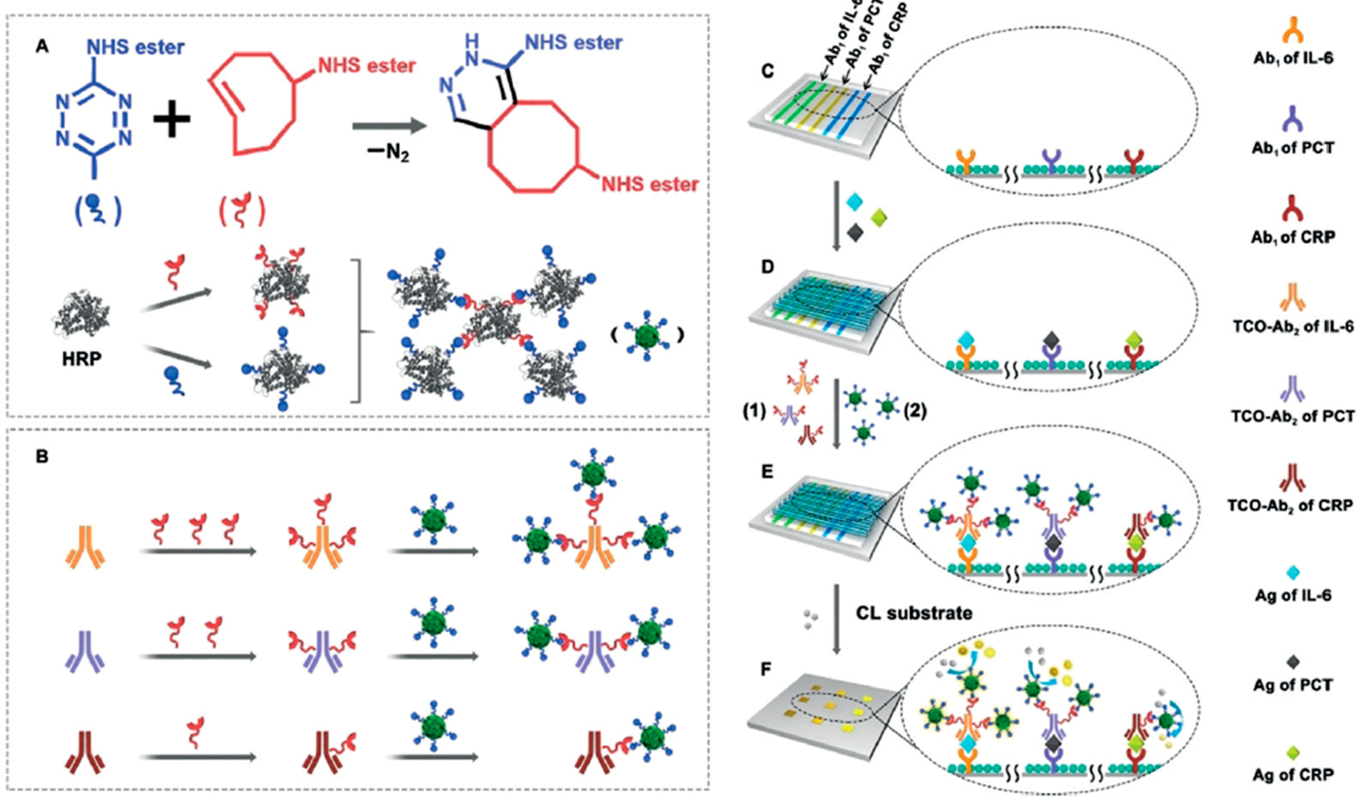
Click-chemistry-mediated assembly of multiple HRPs and detection antibodies for simultaneous multiplexed bioassays with a tunable detection range. Cyclooctene can be quantitatively modified on the detection antibodies to react with Tz-functionalized enzyme assemblies for simultaneous immunoassays of PCT, IL-6, and CRP, each of which requires a different sensitivity. Reproduced from Controllable Assembly of Enzymes for Multiplexed Lab-on-a-Chip Bioassays with a Tunable Detection Range, Xianyu, Y.; Wu, J.; Chen, Y.; Zheng, W.; Xie, M.; Jiang, X. Angew. Chem. Int. Ed., Vol. 57, Issue 25 (ref 52). Copyright 2018 Wiley.
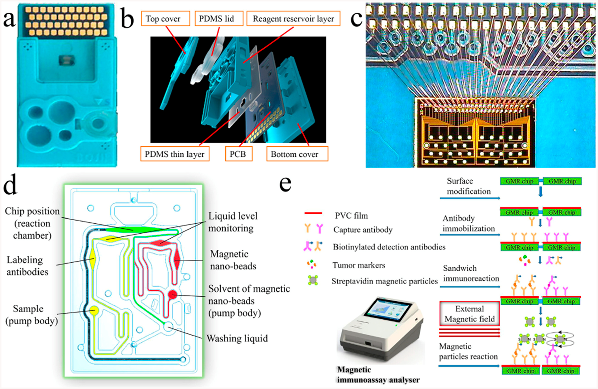
Giant magneto resistance sensor: (a) test card, (b) multilayer structure of the test card, (c) GMR chip and the connection between the GMR chip and PCB, (d) structure of the microchannel system, and (e) reaction process of the GMR multibiomarker immunoassay. Reprinted from Biosensors and Bioelectronics, Vol. 123, Gao, Y.; Huo, W.; Zhang, L.; Lian, J.; Tao, W.; Song, C.; Tang, J.; Shi, S.; Gao, Y. Multiplexed measurement of 12 tumor markers using a GMR multibiomarker immunoassay biosensor, pp. 204–210 (ref 61). Copyright 2019, with permission from Elsevier.
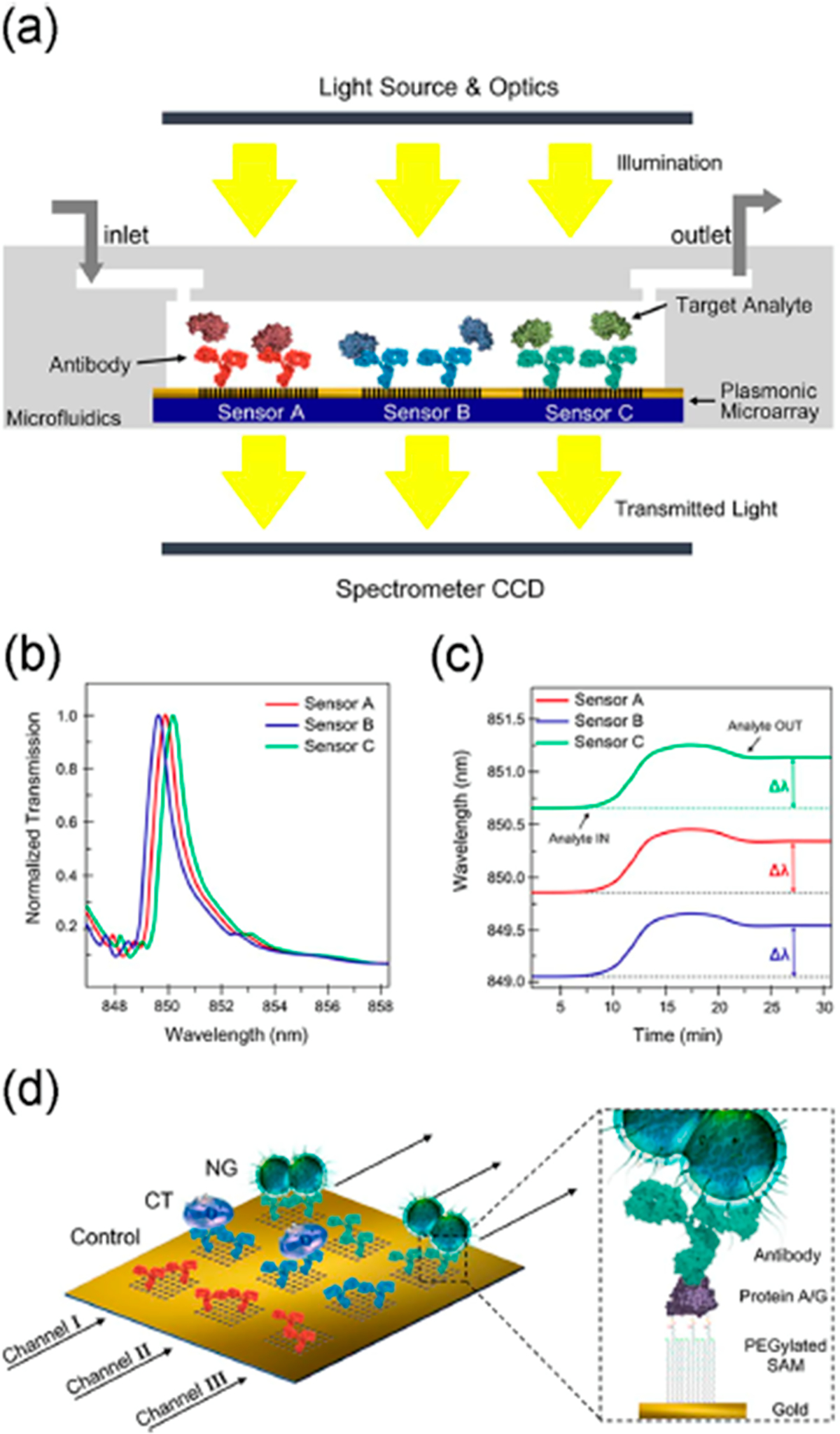
Nanohole biosensor: (a) cross-sectional view, (b) graph illustrating the three EOT spectral peaks acquired simultaneously from the three sensor arrays, (c) graph illustrating the multiplexed real-time monitoring of the EOT wavelength shift, and (d) schematics of sensor surface biofunctionalization. Different nanohole arrays are decorated with different antibodies: anti-Neisseria gonorrheae (NG) (green), anti-Chlamydia trachomatis (CT) (blue), and a control antibody (red). Each microfluidic channel (black arrows) cover three inline sensor arrays. The zoomed-in section illustrates the surface chemistry for the sensor functionalization. Reprinted from Biosensors and Bioelectronics, Vol. 94, Sole, M.; Belushkin, A.; Cavallini, A.; Kebbi-Beghdadi, C.; Greub, G.; Altug, H. Multiplexed nanoplasmonic biosensor for one-step simultaneous detection of Chlamydia trachomatis and Neisseria gonorrheae in urine, pp. 560–567 (ref 69). Copyright 2017, with permission from Elsevier.
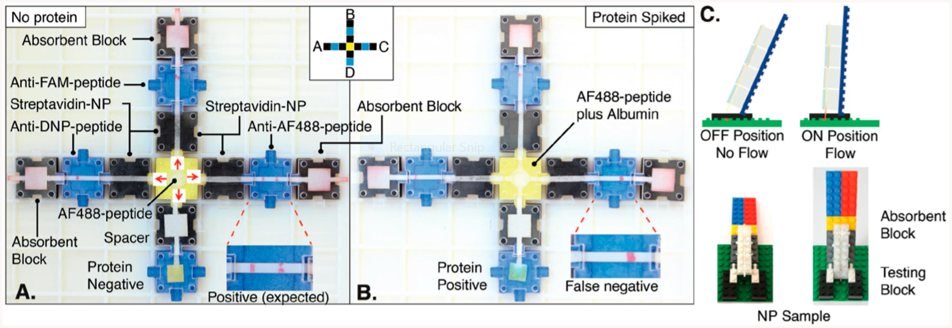
Biochemical breadboard made of a library of linear flow paper fluidic devices that can be put together by the user to construct modular detection systems. Reproduced from Ampli: A Construction Set for Paperfluidic Systems, Phillips, E.A., Young, A.K., Albarran, N., Butler, J., Lujan, K., Hamad-Schifferli, K. and Gomez-Marquez, J. Advanced Healthcare Materials, Vol. 7, Issue 14 (ref 107). Copyright 2018 Wiley.
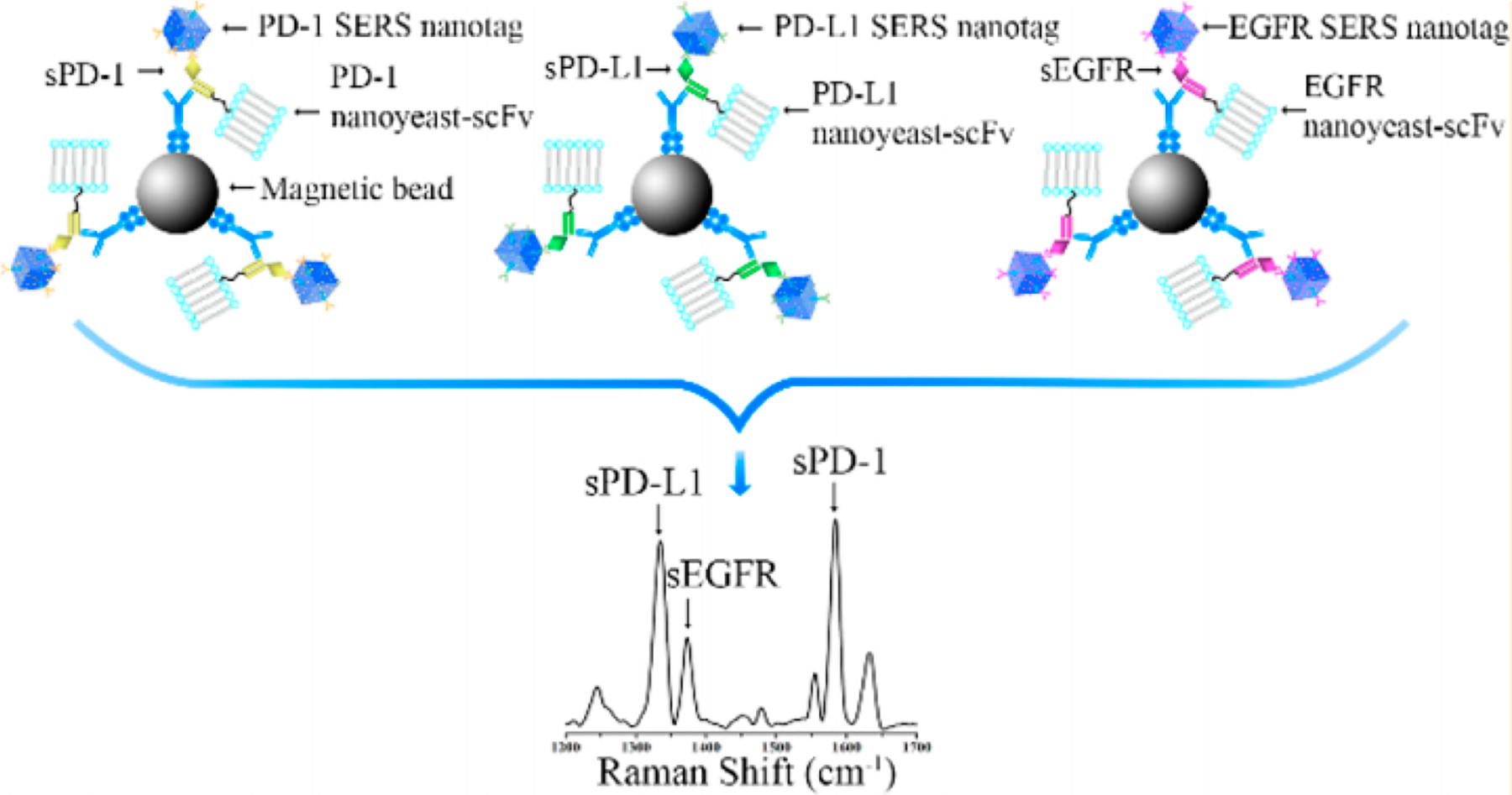
Replacement of antibodies by nanoyeast single chain variable fragments for high sensitivity detection of multiple biomarkers. Reproduced from Li, J.; Wang, J.; Grewal, Y.S.; Howard, C.B.; Raftery, L.J.; Nahler, S.; Wang, Y.; Trau, M. Anal. Chem. 2018, 90, 10377–10384 (ref 116). Copyright 2018 American Chemical Society.
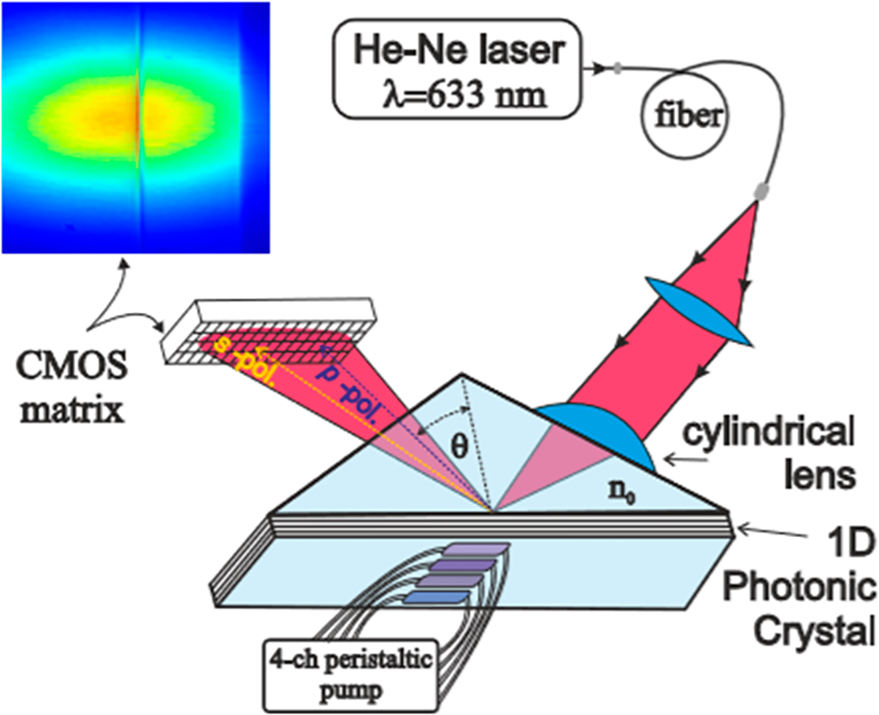
Schematic of the photonic crystal sensor, analogous to that of an SPR chip, and detects signals as molecules on the crystal surface interact with the analytes. Changes in the environment adjacent to the crystal surface changes the intensity of the light with total internal reflection. Reprinted with permission from Petrova, I.; Konopsky, V.; Nabiev, I.; Sukhanova, A. Label-Free Flow Multiplex Biosensing via Photonic Crystal Surface Mode Detection. Sci. Rep. 2019, Vol. 9, 8745 (ref 140). Copyright 2019 Springer Nature.
References
-
- Barry MJN Engl. J. Med 2001, 344, 1373–1377. - PubMed
-
- Ward AM; Catto J; Hamdy F Ann. Clin. Biochem 2001, 38, 633–651. - PubMed
-
- Srinivas PR; Kramer BS; Srivastava S Lancet Oncol. 2001, 2, 698–704. - PubMed
-
- http://www.quansysbio.com/ Last accessed September 1, 2019.
Publication types
MeSH terms
Substances
Grants and funding
LinkOut - more resources
Full Text Sources
