. 2019 Nov 14;20(1):858.
doi: 10.1186/s12864-019-6216-x.

Mouse APOBEC1 cytidine deaminase can induce somatic mutations in chromosomal DNA

Affiliations

Mouse APOBEC1 cytidine deaminase can induce somatic mutations in chromosomal DNA

Vincent Caval et al. BMC Genomics. .

Abstract

Background: APOBEC1 (A1) enzymes are cytidine deaminases involved in RNA editing. In addition to this activity, a few A1 enzymes have been shown to be active on single stranded DNA. As two human ssDNA cytidine deaminases APOBEC3A (A3A), APOBEC3B (A3B) and related enzymes across the spectrum of placental mammals have been shown to introduce somatic mutations into nuclear DNA of cancer genomes, we explored the mutagenic threat of A1 cytidine deaminases to chromosomal DNA.

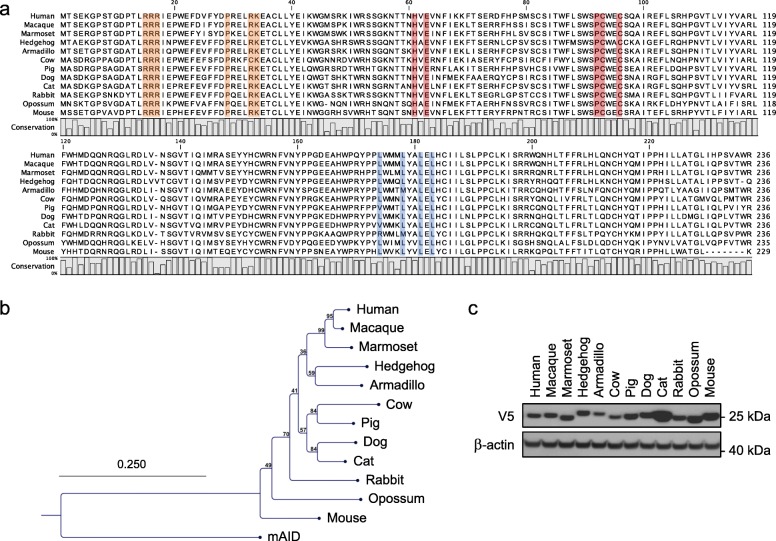
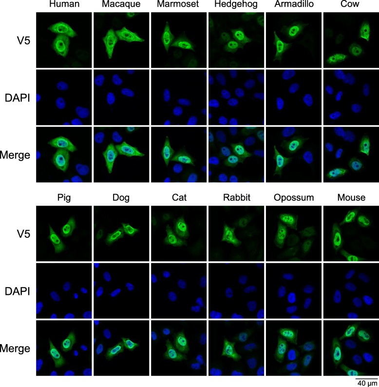
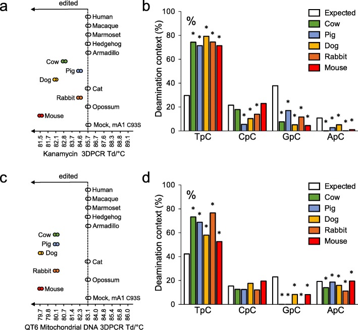
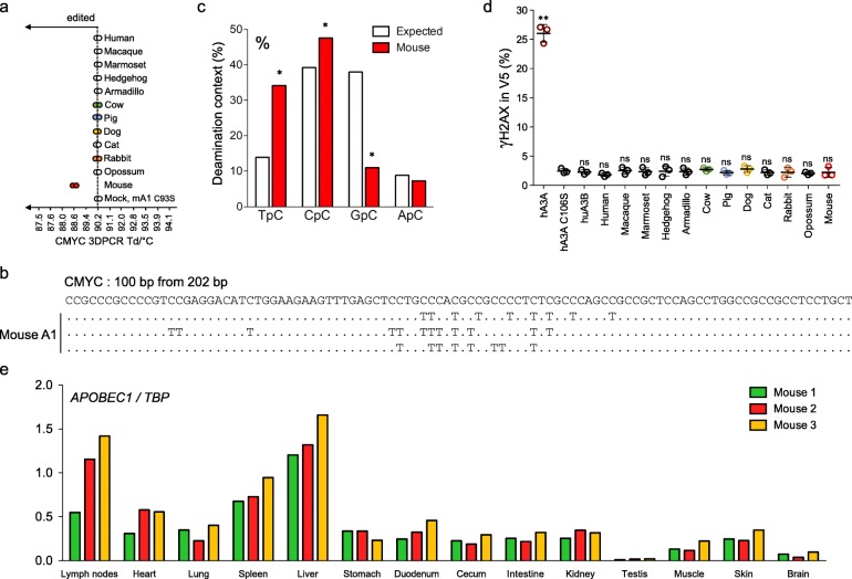
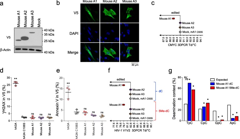
Results: Molecular cloning and expression of various A1 enzymes reveal that the cow, pig, dog, rabbit and mouse A1 have an intracellular ssDNA substrate specificity. However, among all the enzymes studied, mouse A1 appears to be singular, being able to introduce somatic mutations into nuclear DNA with a clear 5'TpC editing context, and to deaminate 5-methylcytidine substituted DNA which are characteristic features of the cancer related mammalian A3A and A3B enzymes. However, mouse A1 activity fails to elicit formation of double stranded DNA breaks, suggesting that mouse A1 possess an attenuated nuclear DNA mutator phenotype reminiscent of human A3B.

Conclusions: At an experimental level mouse APOBEC1 is remarkable among 12 mammalian A1 enzymes in that it represents a source of somatic mutations in mouse genome, potentially fueling oncogenesis. While the order Rodentia is bereft of A3A and A3B like enzymes it seems that APOBEC1 may well substitute for it, albeit remaining much less active. This modifies the paradigm that APOBEC3 and AID enzymes are the sole endogenous mutator enzymes giving rise to off-target editing of mammalian genomes.

Keywords: APOBEC1; Cancer; Cytidine deaminase; Nuclear DNA; Somatic mutations.


Conflict of interest statement

The authors declare that they have no competing interests.

Figures

Fig. 1
Fig. 1
Comparison of APOBEC1 cytidine deaminases. a CLUSTALW alignment of A1 protein sequences. Residues involved in zinc coordination are depicted in red. Residues in orange are part of A1 bipartite nuclear localization signal while those involved in nuclear export of A1 are represented in blue. b Phylogenetic tree of A1 protein sequences constructed using the Neighbor-joining method with the CLC Main Workbench 7.0.2 software. Mouse AID was used to root the tree. Numbers correspond to bootstrap values inferred from 100,000 replicates. c Western blot analysis of V5-tagged A31 proteins in quail QT6 cells. β-actin probing was used as loading control
Fig. 2
Fig. 2
Cellular localization of APOBEC1 cytidine deaminases. Confocal microscopy analysis of V5-tagged A1 proteins in QT6 cells, 24 h post transfection. Nuclei are stained with DAPI
Fig. 3
Fig. 3
APOBEC1 cytidine deaminase activity on plasmid and cytosolic mitochondrial DNA. a Graphical representation of plasmid DNA editing by A1 proteins. The temperature of the DNA products recovered at the lowest Td by kanamycin specific 3DPCR amplification are represented on the gradient. b Dinucleotide analysis of the deamination context performed on plasmid DNA for PCR products retrieved at 84.6 °C. c Graphical representation of cytochrome c mtDNA editing by A1 proteins. The last retrieved bands by cytochrome c specific 3DPCR amplification are represented on the gradient. d Dinucleotide analysis of the deamination context performed on mtDNA for PCR products retrieved at 82.3 °C. Dinucleotide context expected values, based on the dinucleotide composition of DNA sequences are represented by white histograms. * Significant deviation from expected values (χ2-test, P < 0.05)
Fig. 4
Fig. 4
APOBEC1 mediated nuclear DNA editing and damage. a Graphical representation of nuclear DNA editing by A1 proteins. The last positive 3DPCR bands retrieved bands by CMYC specific 3DPCR amplification are represented on the gradient. b Selection of hypermutated CMYC sequences after mouse A1-UGI transfection in QT6 cells for PCR products retrieved at 89.4 °C. c Dinucleotide analysis of mouse A1 deamination context performed on nuclear DNA for PCR products retrieved at 89.4 °C. Dinucleotide context expected values, based on the dinucleotide composition of DNA sequences are represented by white histograms. * Significant deviation from expected values (χ2-test, P < 0.05). d Double strand breaks formation upon A1 transfection in QT6 cells by flow cytometry analysis of γH2AX staining in V5 transfected cells 48 h post-transfection. Human APOBEC3A (hA3A) was used as positive control. Error bars represent the standard deviations from three independent transfections. Differences compared to human APOBEC3A catalytic mutant hA3A C106S were calculated using student t test (** p < 0.01). e APOBEC1 expression in 3 C57/BL6 mice tissues normalized on TBP reference genes
Fig. 5
Fig. 5
APOBEC1 is the only mouse APOBEC cytidine deaminase capable of mutating nuclear and 5-methylcytidine containing DNA. a Western blot analysis of V5-tagged mouse APOBEC cytidine deaminases in quail QT6 cells. β-actin probing was used as loading control. b Confocal microscopy analysis of V5-tagged mouse APOBEC cytidine deaminases in QT6 cells, 24 h post transfection. Nuclei are stained with DAPI. c Graphical representation of nuclear DNA editing by mouse APOBEC cytidine deaminases. The last retrieved bands by CMYC specific 3DPCR amplification are represented on the gradient. d Double strand breaks formation upon mouse APOBEC cytidine deaminases transfection in QT6 cells by flow cytometry analysis of γH2AX staining in V5 transfected cells 48 h post-transfection. Human APOBEC3A (hA3A) was used as positive control. Error bars represent the standard deviations of three independent transfections. Differences compared to human APOBEC3A catalytic mutant hA3A C106S were calculated using student t test (** P < 0.01). e Annexin V staining of apoptosis upon mouse APOBEC cytidine deaminases transfection in HeLa cells by flow cytometry analysis in V5 transfected cells 36 h post-transfection. Differences compared to human APOBEC3A catalytic mutant hA3A C106S were calculated using student t test (** P < 0.01). f Graphical representation of HIV-1 V1 V2 specific 3D-PCR amplification after QT6 transfections with APOBEC cytidine deaminases plasmids along with a cytidine (dC) or 5-methylcytidine (5Me-dC) containing HIV-1 env DNA. g Dinucleotide analysis of mouse A1 deamination context performed on HIV-1 V1 V2 sequences obtained at 81.2 °C from DNA containing either cytidine (dC) or 5-methylcytidine (5Me-dC). Dinucleotide context expected values, based on the dinucleotide composition of DNA sequences are represented by white histograms. * Significant deviation from expected values (χ2-test, P < 0.05)

References

    1. Navaratnam N, Morrison JR, Bhattacharya S, Patel D, Funahashi T, Giannoni F, et al. The p27 catalytic subunit of the apolipoprotein B mRNA editing enzyme is a cytidine deaminase. J Biol Chem. 1993;268:20709–20712. - PubMed
    1. Teng B, Burant CF, Davidson NO. Molecular cloning of an apolipoprotein B messenger RNA editing protein. Science. 1993;260:1816–1819. doi: 10.1126/science.8511591. - DOI - PubMed
    1. Blanc V, Davidson NO. APOBEC-1-mediated RNA editing. Wiley Interdiscip Rev Syst Biol Med. 2010;2:594–602. doi: 10.1002/wsbm.82. - DOI - PMC - PubMed
    1. Lellek H, Kirsten R, Diehl I, Apostel F, Buck F, Greeve J. Purification and molecular cloning of a novel essential component of the apolipoprotein B mRNA editing enzyme-complex. J Biol Chem. 2000;275:19848–19856. doi: 10.1074/jbc.M001786200. - DOI - PubMed
    1. Mehta A, Kinter MT, Sherman NE, Driscoll DM. Molecular cloning of apobec-1 complementation factor, a novel RNA-binding protein involved in the editing of apolipoprotein B mRNA. Mol Cell Biol. 2000;20:1846–1854. doi: 10.1128/MCB.20.5.1846-1854.2000. - DOI - PMC - PubMed