. 2019 Nov 14;9(1):16742.
doi: 10.1038/s41598-019-52966-0.

Performance of five automated white matter hyperintensity segmentation methods in a multicenter dataset

Collaborators, Affiliations

Performance of five automated white matter hyperintensity segmentation methods in a multicenter dataset

Rutger Heinen et al. Sci Rep. .

Abstract

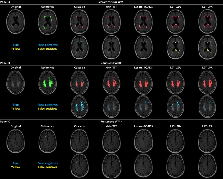
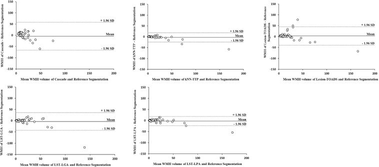
White matter hyperintensities (WMHs) are a common manifestation of cerebral small vessel disease, that is increasingly studied with large, pooled multicenter datasets. This data pooling increases statistical power, but poses challenges for automated WMH segmentation. Although there is extensive literature on the evaluation of automated WMH segmentation methods, such evaluations in a multicenter setting are lacking. We performed WMH segmentations in sixty patients scanned on six different magnetic resonance imaging (MRI) scanners (10 patients per scanner) using five freely available and fully-automated WMH segmentation methods (Cascade, kNN-TTP, Lesion-TOADS, LST-LGA and LST-LPA). Different MRI scanner vendors and field strengths were included. We compared these automated WMH segmentations with manual WMH segmentations as a reference. Performance of each method both within and across scanners was assessed using spatial and volumetric correspondence with the reference segmentations by Dice's similarity coefficient (DSC) and intra-class correlation coefficient (ICC) respectively. We found the best performance, both within and across scanners, for kNN-TTP, followed by LST-LPA and LST-LGA, with worse performance for Lesion-TOADS and Cascade. Our findings can serve as a guide for choosing a method and also highlight the importance to further improve and evaluate consistency of methods in a multicenter setting.


Conflict of interest statement

The authors declare no competing interests.

Figures

Figure 1
Figure 1
WMH segmentations of the methods regarding periventricular, confluent and punctuate WMHs. Example of WMH segmentations for a subject (subject A) with predominantly periventricular WMHs (panel A), a subject (subject B) with large confluent WMHs (panel B) and a subject (subject C) with predominantly punctuate WMHs (panel C). Top rows panels (A–C) original FLAIR scan and WMH reference segmentation (green) and WMH segmentations of all methods (red) are shown. Bottom rows panels (A–C) false negative voxels are shown in blue; false positive voxels are shown in yellow.
Figure 2
Figure 2
Bland Altman plots comparing WMH volume of each method versus the WMH volume of the reference segmentations. X-axis: mean WMH volume (in mL) of the automated and reference segmentations. Y-axis: difference (in mL) in WMH volume between the automated and reference segmentations. The lower (−1.96 SD) and upper (+1.96 SD) limits of agreement (dashed lines) and mean (straight line) are shown. A narrow width of the limits of agreement reflects a small amount of variation between the measurements of the reference and automated WMH segmentations. A positive difference on the y-axis is seen when WMH volume as measured by the automated method was larger than the reference WMH volume (i.e. overestimation). A negative difference on the y-axis is seen when WMH volume as measured by the automated method was smaller than the reference WMH volume (i.e. underestimation).

References

    1. Carrillo MC, Bain LJ, Frisoni GB, Weiner MW. Worldwide Alzheimer’s disease neuroimaging initiative. Alzheimers. Dement. 2012;8:337–42. doi: 10.1016/j.jalz.2012.04.007. - DOI - PubMed
    1. Williamson, J. D. et al. The Action to Control Cardiovascular Risk in Diabetes Memory in Diabetes Study (ACCORD-MIND): Rationale, Design, and Methods. Am. J. Cardiol. 99 (2007). - PubMed
    1. Mueller SG, et al. Ways toward an early diagnosis in Alzheimer’s disease: The Alzheimer’s Disease Neuroimaging Initiative (ADNI) Alzheimer’s Dement. 2005;1:55–66. doi: 10.1016/j.jalz.2005.06.003. - DOI - PMC - PubMed
    1. De Guio F, et al. Reproducibility and variability of quantitative magnetic resonance imaging markers in cerebral small vessel disease. J. Cereb. Blood Flow Metab. 2016;36:1319–1337. doi: 10.1177/0271678X16647396. - DOI - PMC - PubMed
    1. Caligiuri ME, et al. Automatic Detection of White Matter Hyperintensities in Healthy Aging and Pathology Using Magnetic Resonance Imaging: A Review. Neuroinformatics. 2015;13:261–276. doi: 10.1007/s12021-015-9260-y. - DOI - PMC - PubMed

Publication types

LinkOut - more resources