. 2019 Nov 14;9(1):16825.
doi: 10.1038/s41598-019-53259-2.

Identification of candidate molecular targets of the novel antineoplastic antimitotic NP-10

Affiliations

Identification of candidate molecular targets of the novel antineoplastic antimitotic NP-10

Takuya Yokoyama et al. Sci Rep. .

Abstract

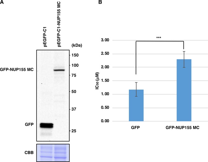
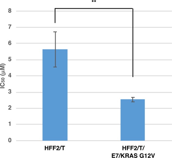
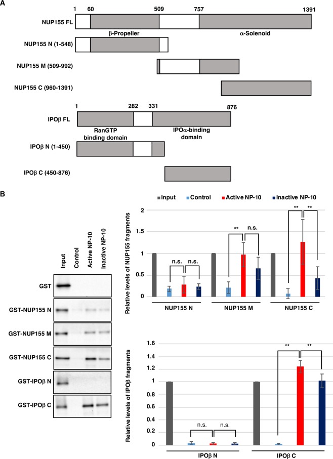
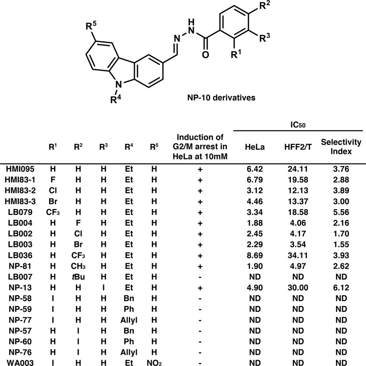
We previously reported the identification of a novel antimitotic agent with carbazole and benzohydrazide structures: N'-[(9-ethyl-9H-carbazol-3-yl)methylene]-2-iodobenzohydrazide (code number NP-10). However, the mechanism(s) underlying the cancer cell-selective inhibition of mitotic progression by NP-10 remains unclear. Here, we identified NP-10-interacting proteins by affinity purification from HeLa cell lysates using NP-10-immobilized beads followed by mass spectrometry. The results showed that several mitosis-associated factors specifically bind to active NP-10, but not to an inactive NP-10 derivative. Among them, NUP155 and importin β may be involved in NP-10-mediated mitotic arrest. Because NP-10 did not show antitumor activity in vivo in a previous study, we synthesized 19 NP-10 derivatives to identify more effective NP-10-related compounds. HMI83-2, an NP-10-related compound with a Cl moiety, inhibited HCT116 cell tumor formation in nude mice without significant loss of body weight, suggesting that HMI83-2 is a promising lead compound for the development of novel antimitotic agents.


Conflict of interest statement

The authors declare no competing interests.

Figures

Figure 1
Figure 1
Overexpression of NUP155 MC (aa 748–1391) confers partial resistance to NP-10 cytotoxicity in HeLa cells. (A) HeLa cells were transfected with the indicated expression vectors for 48 h and immunoblotted with anti-GFP antibody. Proteins were stained with Coomassie Brilliant Blue (CBB) to check equal loading. (B) HeLa cells were transfected with the indicated expression vectors for 24 h and subjected to colony formation assays in the presence of NP-10 at various concentrations. IC50 values were calculated. Data represent the mean ± SD from nine independent experiments. ***p < 0.001 (two-tailed Student’s t-test).
Figure 2
Figure 2
Transformed HFF2/T/E7/KRAS G12V cells are hypersensitive to NP-10 compared with parental HFF2/T cells. Parental HFF2/T and HFF2/T/E7/KRAS G12V cells were subjected to colony formation assays in the presence of NP-10 at various concentrations, and the IC50 values were calculated. Data represent the mean ± SD from multiple independent experiments (n = 4 for HFF2/T, n = 3 for HFF2/T/E7/KRAS G12V). **p < 0.01 (two-tailed Student’s t-test).
Figure 3
Figure 3
Pull-down assay with biotin-conjugated compounds. (A) Chemical structures of biotinylated compounds. O-PEGylated NP-10 (R1 = I, R2 = H) or NP-14 (R1 = H, R2 = I) linked to PEGylated biotin via the 1,2,3-triazole moiety (upper panel) and N-PEGylated NP-10 or NP-14 linked to PEGylated biotin via the 1,2,3-triazole moiety (lower panel) are shown. (B) O-PEGylated, but not N-PEGylated, NP-10 and NP-14 (without 1,2,3-triazole and PEGylated biotin) induce G2/M arrest in HeLa cells. HeLa cells were treated with vehicle (DMSO) or the indicated compounds (10 μM) for 24 h. After staining with propidium iodide, cell cycle distribution was analyzed by flow cytometry (upper panels). In vitro inhibition of HeLa cell growth by the compounds was also investigated (lower panel). The IC50 (50% inhibition concentration) values were estimated. Data represent the mean from two independent experiments. (C) HeLa cell lysates were incubated with the O-biotinylated (active) or N-biotinylated (inactive) NP-10 beads immobilized by biotin-avidin interaction. The pulled-down proteins were analyzed by SDS-PAGE and Coomassie Brilliant Blue (CBB) staining. The gel was cut into eight fractions as shown on the right. Proteins in each gel fraction were identified by mass spectrometry analysis.
Figure 4
Figure 4
Overexpression of GFP-IPOβ and silencing of NUP155 induce mitotic arrest. (A) HeLa cells were co-transfected with the indicated expression vectors and siRNAs for 48 h, and immunoblotted with the indicated antibodies. (B) HeLa cells transfected as in (A) were stained with DAPI and analyzed by fluorescence microscopy. Representative images of mitotic cells are shown. Scale bar, 10 μm. Percentages of mitotic cells with GFP signals are depicted. *p < 0.05, **p < 0.01, ***p < 0.001 (χ2-test). Similar results were obtained in two independent experiments.
Figure 5
Figure 5
HMI83-2 suppresses the growth of HCT116 tumors in nude mice. (A) Chemical structure of HMI83-2. (B) HCT116 cells were subcutaneously inoculated into BALB/c nu/nu nude mice. The mice were treated with 100 mg/kg of HMI83-2 or control vehicle (n = 7, n = 6 respectively), as described in Materials and Methods. The mean tumor volumes (with SDs) are shown. Arrows represent the days of drug administration. *p < 0.05 (compared with vehicle; two-tailed Student’s t-test). (C) Kaplan–Meier curves obtained with the experiments shown above. *p < 0.05 (compared with vehicle; log-rank test). (D) Changes in body weight of the nude mice treated with HMI83-2 or control vehicle are shown.
Figure 6
Figure 6
Silencing of NUP155, a potential target of NP-10, induces mitotic arrest. (A) HeLa cell lysates were incubated with avidin beads on which the indicated compounds were immobilized. The bound proteins and 5.4% of the input were analyzed by immunoblotting with the indicated antibodies. (B) HeLa cells were transfected with control (mixture of siGFP and siLuci) or NUP155-targeting (siNUP155-1 or siNUP155-2) siRNAs for 48 h. Whole cell extracts were analyzed by immunoblotting with the indicated antibodies. Phospho-vimentin (Ser55), a target of Cdk1 kinase, was used as a mitotic marker. Proteins were stained with Coomassie Brilliant Blue (CBB) to check equal loading. (C) HeLa cells treated as above were stained with propidium iodide and subjected to flow cytometry.
Figure 7
Figure 7
Identification of NP-10-binding domains for IPOβ and NUP155. (A) Schematic representation of full-length NUP155 and IPOβ (FL) and their truncated mutants. The N-terminal and C-terminal parts of NUP155 have predicted β-propeller and α-solenoid structures, respectively. The N-terminal and C-terminal parts of IPOβ are RanGTP and IPOα binding domains, respectively. (B) GST, GST-IPOβ mutants, or NUP155 mutants were incubated with active or inactive NP-10-immobilized beads (through biotin-avidin interaction). The pulled-down proteins, 3.5% of the input for GST-NUP155 mutants, and 5.8% of the input for GST and GST-IPOβ mutants were analyzed by immunoblotting with anti-GST antibody. Biotin-conjugated PEG linker was used as a negative control. The signal intensities of the bands were quantified and shown with the signal intensities of inputs set as 1. The mean ± SD is shown (n = 3 for NUP155 N and IPOβ N; n = 4 for NUP155 M; n = 5 for NUP155C and IPOβ C). **p < 0.01, n.s., not significant (Tukey-Kramer test).
Figure 8
Figure 8
Growth inhibitory activities of various NP-10-related compounds. Chemical structures of NP-10 derivatives and their growth inhibitory activities. The IC50 unit was μM. The selectivity index is the ratio between the IC50 for HFF2/T cells and the IC50 for HeLa cells. ND, not determined.

References

    1. Chan K-S, Koh C-G, Li H-Y. Mitosis-targeted anti-cancer therapies: where they stand. Cell Death Dis. 2012;3:e411. doi: 10.1038/cddis.2012.148. - DOI - PMC - PubMed
    1. Kavallaris M. Microtubules and resistance to tubulin-binding agents. Nat. Rev. Cancer. 2010;10:194–204. doi: 10.1038/nrc2803. - DOI - PubMed
    1. Harrison MR, Holen KD, Liu G. Beyond taxanes: a review of novel agents that target mitotic tubulin and microtubules, kinases, and kinesins. Clin. Adv. Hematol. Oncol. 2009;7:54–64. - PMC - PubMed
    1. Jordan MA, Wilson L. Microtubules as a target for anticancer drugs. Nat. Rev. Cancer. 2004;4:253–65. doi: 10.1038/nrc1317. - DOI - PubMed
    1. Ohira M, et al. A novel anti-microtubule agent with carbazole and benzohydrazide structures suppresses tumor cell growth in vivo. Biochim. Biophys. Acta - Gen. Subj. 2015;1850:1676–1684. doi: 10.1016/j.bbagen.2015.04.013. - DOI - PubMed

Publication types

MeSH terms