. 2019 Nov 14;9(1):16829.
doi: 10.1038/s41598-019-53133-1.

An Improved Free Energy Perturbation FEP+ Sampling Protocol for Flexible Ligand-Binding Domains

Affiliations

An Improved Free Energy Perturbation FEP+ Sampling Protocol for Flexible Ligand-Binding Domains

Filip Fratev et al. Sci Rep. .

Abstract

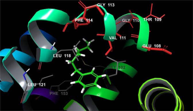
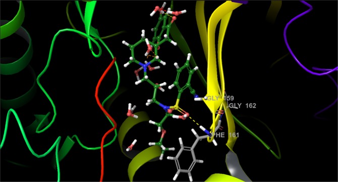
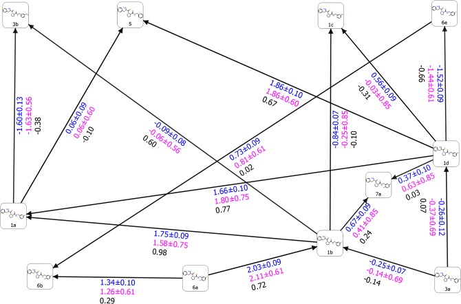
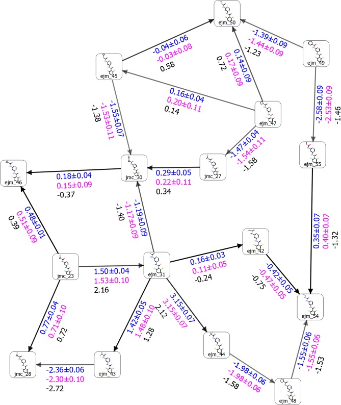
Recent improvements to the free energy perturbation (FEP) calculations, especially FEP+ , established their utility for pharmaceutical lead optimization. Herein, we propose a modified version of the FEP/REST (i.e., replica exchange with solute tempering) sampling protocol, based on detail studies on several targets by probing a large number of perturbations with different sampling schemes. Improved FEP+ binding affinity predictions for regular flexible-loop motions and considerable structural changes can be obtained by extending the prior to REST (pre-REST) sampling time from 0.24 ns/λ to 5 ns/λ and 2 × 10 ns/λ, respectively. With this new protocol, much more precise ∆∆G values of the individual perturbations, including the sign of the transformations and decreased error were obtained. We extended the REST simulations from 5 ns to 8 ns to achieve reasonable free energy convergence. Implementing REST to the entire ligand as opposed to solely the perturbed region, and also some important flexible protein residues (pREST region) in the ligand binding domain (LBD) has considerably improved the FEP+ results in most of the studied cases. Preliminary molecular dynamics (MD) runs were useful for establishing the correct binding mode of the compounds and thus precise alignment for FEP+ . Our improved protocol may further increase the FEP+ accuracy.


Conflict of interest statement

The authors declare no competing interests.

Figures

Figure 1
Figure 1
A flowchart which represents the steps of suggested new sampling protocol and comparison with the default one.
Figure 2
Figure 2
Structure of LBD of T4 lysozyme (PDB id 4W52) bond with Hexylbenzene. Residues from F-Helix (colored in red) are flexible and determine the LBD in an open, closed and intermediate state.
Figure 3
Figure 3
(A) Free energy output maps obtained via the 2 × 10 ns pre-REST FEP+ sampling protocol for all T4 lysozyme L99A test set of ligands and (B) for the compounds for which experimental ∆∆G values are available and the pREST protocol was also applied. Black, blue, and red numbers indicate the experimental (∆∆Gexp), calculated Bennett (∆∆Gpred), and cycle closure (∆∆Gpredc) free energies of binding, respectively.
Figure 4
Figure 4
Structure of LBD of AKT1 (PDB id 3QKK) bond with ligand 18. The substrate peptide and F-loop are colored in red and yellow, respectively. The H-bonds are shown in yellow dot lines.
Figure 5
Figure 5
Free energy output maps obtained via the 5 ns pre-REST FEP+ sampling protocol for (A) selected small and (B) full test set of AKT1 ligands. Black, blue, and red numbers indicate the experimental ∆∆Gexp, calculated Bennett (∆∆Gpred), and cycle closure (∆∆Gpredc) free energies of binding, respectively.
Figure 6
Figure 6
Structure of LBD of THR (PDB id 2ZFF) bond with ligand 1a. Ligand is rendered as green stick model and protein rendered as cartoon model. The H-bonds are shown in yellow dot lines.
Figure 7
Figure 7
Free energy output map obtained via the 5 ns pre-REST FEP+ sampling protocol for the selected test set of THR ligands. Black, blue, and red numbers indicate the experimental (∆∆Gexp), calculated Bennett (∆∆Gpred), and cycle closure (∆∆Gpredc) free energies of binding, respectively.
Figure 8
Figure 8
Structure of LBD of TYK2 (PDB id: 4GIH) bond with ligand ejm_46. The H-bonds are shown in yellow dot lines.
Figure 9
Figure 9
Free energy output map obtained via the 5 ns pre-REST FEP+ sampling protocol for the selected test set of TYK2 ligands. Black, blue, and red numbers indicate the experimental (∆∆Gexp), calculated Bennett (∆∆Gpred), and cycle closure (∆∆Gpredc) free energies of binding, respectively.

References

    1. Paul SM, et al. How to improve rd productivity: the pharmaceutical industry’s grand challenge. Nat. Rev. Drug Discov. 2010;9:203. doi: 10.1038/nrd3078. - DOI - PubMed
    1. Waring MJ, et al. An analysis of the attrition of drug candidates from four major pharmaceutical companies. Nat. Rev. Drug Discov. 2015;14:475. doi: 10.1038/nrd4609. - DOI - PubMed
    1. Wang L, et al. Accurate and reliable prediction of relative ligand binding potency in prospective drug discovery by way of a modern free-energy calculation protocol and force field. J. Am. Chem. Soc. 2015;137:2695–2703. doi: 10.1021/ja512751q. - DOI - PubMed
    1. Steinbrecher TB, et al. Accurate binding free energy predictions in fragment optimization. J. Chem. Inf. Model. 2015;55:2411–2420. doi: 10.1021/acs.jcim.5b00538. - DOI - PubMed
    1. Kuhn B, et al. Prospective evaluation of free energy calculations for the prioritization of cathepsin l inhibitors. J. Medicinal Chem. 2017;60:2485–2497. doi: 10.1021/acs.jmedchem.6b01881. - DOI - PubMed

Publication types