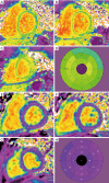
Multi-modal imaging of the pediatric heart transplant recipient
- PMID: 31728325
- PMCID: PMC6825966
- DOI: 10.21037/tp.2019.08.04
Multi-modal imaging of the pediatric heart transplant recipient
Abstract
The assessment of pediatric patients after orthotropic heart transplantation (OHT) relies heavily on non-invasive imaging. Because of the potential risks associated with cardiac catheterization, expanding the role of non-invasive imaging is appealing. Echocardiography is fast, widely available, and can provide an accurate assessment of chamber sizes and function. Advanced echocardiographic methods, such as myocardial deformation, have potential to assess for acute rejection or cardiac allograft vasculopathy (CAV). While not currently part of routine care, cardiac magnetic resonance imaging (CMR) and computed tomography may potentially aid in the detection of graft complications following OHT. In particular, CMR tissue characterization holds promise for diagnosing rejection, while quantitative perfusion and myocardial late gadolinium enhancement may have a role in the detection of CAV. This review will evaluate standard and novel methods for non-invasive assessment of pediatric patients after OHT.
Keywords: Cardiac imaging; cardiac computed tomography; cardiac magnetic resonance imaging (CMR); echocardiography; orthotopic heart transplantation (OHT).
2019 Translational Pediatrics. All rights reserved.
Conflict of interest statement
Conflicts of Interest: The authors have no conflicts of interest to declare.
Figures


References
-
- Khush KK, Cherikh WS, Chambers DC, et al. The International Thoracic Organ Transplant Registry of the International Society for Heart and Lung Transplantation: Thirty-fifth Adult Heart Transplantation Report-2018; Focus Theme: Multiorgan Transplantation. J Heart Lung Transplant 2018;37:1155-68. 10.1016/j.healun.2018.07.022 - DOI - PubMed
-
- Rossano JW, Cherikh WS, Chambers DC, et al. The International Thoracic Organ Transplant Registry of the International Society for Heart and Lung Transplantation: Twenty-first pediatric heart transplantation report-2018; Focus theme: Multiorgan Transplantation. J Heart Lung Transplant 2018;37:1184-95. 10.1016/j.healun.2018.07.018 - DOI - PubMed
Publication types
Grants and funding
LinkOut - more resources
Full Text Sources
