. 2020 Jan 10;48(1):359-372.
doi: 10.1093/nar/gkz1065.

RbfA and IF3 couple ribosome biogenesis and translation initiation to increase stress tolerance

Affiliations

RbfA and IF3 couple ribosome biogenesis and translation initiation to increase stress tolerance

Indra Mani Sharma et al. Nucleic Acids Res. .

Abstract

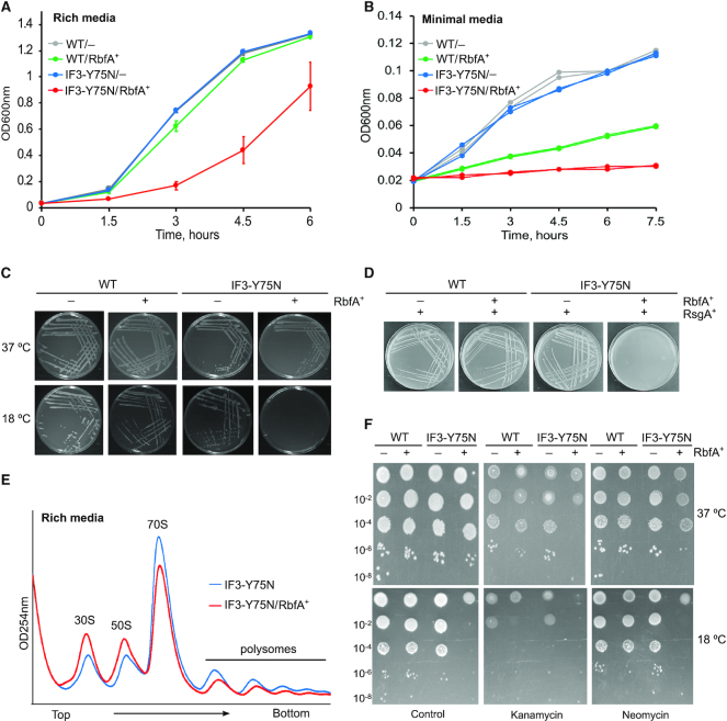
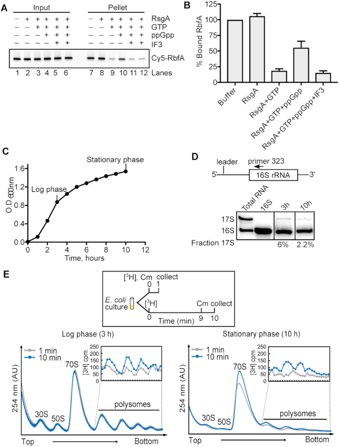
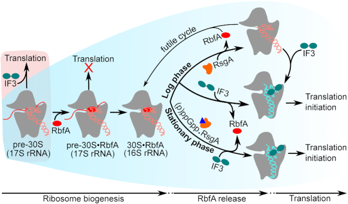
Bacterial ribosome biogenesis and translation occur in the same cellular compartment. Therefore, a biochemical gate-keeping step is required to prevent error-prone immature ribosomes from engaging in protein synthesis. Here, we provide evidence for a previously unknown quality control mechanism in which the abundant ribosome assembly factor, RbfA, suppresses protein synthesis by immature Escherichia coli 30S subunits. After 30S maturation, RbfA is displaced by initiation factor 3 (IF3), which promotes translation initiation. Genetic interactions between RbfA and IF3 show that RbfA release by IF3 is important during logarithmic growth as well as during stress encountered during stationary phase, low nutrition, low temperature, and antibiotics. By gating the transition from 30S biogenesis to translation initiation, RbfA and IF3 maintain the fidelity of bacterial protein synthesis.


Figures

Figure 1.
Figure 1.
Full-length IF3 is required for RbfA release from 30S ribosomes. (A) Results of pelleting assay showing the release of Cy5-RbfA from 30S•Cy5-RbfA complexes in the presence of initiation factors. Only IF3 was able to displace RbfA from 30S subunits (lane 10). A complex of mature 30S (200 nM) and Cy5-RbfA (400 nM) was formed and unbound Cy5-RbfA was removed by filtration (input) before complexes were challenged with initiation factors (4 μM). Proteins were resolved by 4–20% SDS PAGE. Top panel, Cy5-RbfA fluorescence; bottom panel, Coomassie stain. Initiation factors (input) are indicated with black dots. (B) Pelleting assay showing that a non-binding IF3 mutant (IF3-K110L) cannot release of RbfA from 30S subunits (lane 9). RsgA (500 nM) and GTP (5 μM) was used as a positive control (lane 10). *RbfA (lanes 1 and 6); Cy5-RbfA only. (C) Results of pelleting assay showing that a mutation in the linker region of IF3 (IF3-Y75N) reduces Cy5-RbfA release. (D) Separated N- and C-terminal domains of IF3 were used alone or in combination. (E) Fraction of bound Cy5-RbfA in pellets from panels (A)–(D). Bars indicate mean and s.d., n = 3 independent trials. Dotted line indicates ∼5% Cy5-RbfA background in reactions lacking 30S subunits.
Figure 2.
Figure 2.
IF3 cannot release RbfA from immature pre-30S subunits. (A) Composition of pre-30S ΔrbfA particles. Left, 1.5% agarose-TAE gel showing the rRNA profile. Fraction 2 containing >90% 17S pre-rRNA (pre-30S) was used for further assays. Right, 4–20% SDS PAGE comparing proteins from mature 30S and pre-30S ΔrbfA particles. Proteins S1, S2 and S3 are missing in the pre-30S particles. The identity of the extra band (*) is not known. (B) Pelleting assay; Cy5-RbfA (400 nM) was complexed with mature 30S or pre-30S subunits (200 nM) and challenged with IF3 (4 μM) as in Figure 1. (C) Kinetics of Cy5-RbfA dissociation from pre-30S and 30S subunits. Top, with or without 4 μM IF3; bottom, with or without 4 μM unlabeled RbfA. After excess Cy5-RbfA was removed from Cy5-RbfA•30S complexes by filtration (‘0 min’), complexes were filtered a second time to remove free Cy5-RbfA after an additional 20–60 min at 37°C. The 0 min samples that were filtered once contain ∼30% more Cy5-RbfA than the samples that were filtered twice. (D) Binding of Cy5-RbfA and Cy3-IF3 to pre-30S or 30S subunits, as in C. Top panel, Cy5-RbfA; middle panel, Cy3-IF3; lower panel, Coomassie stain. *IF3 (lanes 1 and 8); Cy3-IF3 only. Average fold change in bound IF3 over *IF3 background (lane 8) was 1.9 ± 0.15 in lanes 10 and 11 and 2.2 ± 0.9 in lanes 13 and 14; n = 2. Added IF3 was a mixture of 80% unlabeled IF3 and 20% Cy3-IF3. Cy5-RbfA was scanned with 600 PMT voltage, whereas Cy3-IF3 (Input) with 400 PMT voltage and Cy3-IF3 (retentate) with 500 PMT voltage. (E) Anti-IF3 western blot showing the presence of IF3 in the pre-30S and 30S fractions from BX41 (ΔrbfA) and BW25113 (WT) compared to ΔrbfA and WT lysates and purified IF3 (control).
Figure 3.
Figure 3.
Methylation of 30S subunits is important for RbfA release. (A) Methylation of 16S A1519 in pre-30S and 30S subunits was measured by primer extension (see Materials and Methods). Pre-30S subunits from BX41 (ΔrbfA) were methylated by KsgA (lane 4); nm30S subunits from ΔksgA cells (JW0050-3) and TPR201 cells bearing an inactive ksgA allele were unmethylated (lanes 6 and 7). (B) Pelleting assay showing that neither IF3 (top) nor RsgA (bottom) promote Cy5-RbfA release from nm30SΔksgA subunits and promote only partial release from nm30STPR201 subunits. (C) % Bound RbfA relative to buffer control as in panel B; mean and s.d.; n = 2. (D) Native PAGE showing that Cy5-RbfA and Cy3-IF3 can bind pre-30S or 30S subunits simultaneously. 30S complexes were purified from the strains shown and incubated with Cy5-RbfA and Cy3-IF3 before native PAGE. Panels show the same gel scanned with Cy3 excitation (bottom), Cy5 excitation (middle), and FRET to Cy5 upon Cy3 excitation (top). The FRET efficiencies are indicated at the bottom.
Figure 4.
Figure 4.
Translation initiation requires RbfA release from 30S subunits. (A) In vitro translation by pre-30S (methylated), nm30S (unmethylated) and mature 30S subunits in the presence of RbfA or RbfA plus IF3. Top, average and s.d., n = 2, relative to no RbfA controls for each complex. Bottom, 35S-labeled DHFR product. Immature or unmethylated subunits are active in protein synthesis but are inhibited by RbfA. Mature 30S subunits were unaffected by RbfA, presumably because RbfA was removed by IF3. (B) 16S rRNA processing. Binding of anti-sense oligomers to RNA from 30S complexes was detected by native 4% PAGE.
Figure 5.
Figure 5.
Genetic interaction between RbfA and IF3. (A) Growth of E. coli strains in rich LB media at 37°C (OD600nm). WT, parental; IF3-Y75N, infC362; –, empty vector; and RbfA+, + p15BHA (RbfA overexpression). Mean and s.d., n = 4 biological replicates. (B) Growth of strains in (A) in minimal MOPS media (pH 7.2) supplemented with 0.4% glucose at 37°C; n = 2. (C) Growth of strains as in (A) on LB agar media plates at 37°C and 18°C; n = 3. (D) WT and IF3-Y75N cells harboring empty vector or p15BHA were transformed with pD421-rsgA for RsgA overexpression. Plates were incubated overnight at 37°C. Single colonies were subsequently streaked on a fresh plate. No transformation was observed for the IF3-Y75N/RbfA+/RsgA+ strain, n = 2. (E) Polysome profiles of strains infC362 (blue) and infC362/p15BHA (red), grown under similar conditions as (A) and collected at OD600nm ∼0.2. Experiment was performed in duplicate. (F) Serial dilutions of the indicated strains were spotted on LB-agar containing sublethal concentrations of kanamycin (2 μg/ml) or neomycin (2 μg/ml). Ampicillin (100 μg/ml) was added to all plates to maintain the plasmid; n = 2.
Figure 6.
Figure 6.
IF3 releases RbfA from 30S ribosomes during stationary phase. (A) Pelleting assay showing that RbfA release by 200 nM RsgA, 50 μM GTP is inhibited by 1.5 mM ppGpp (lane 10). 5 μM IF3 can release RbfA from 30S subunits under this condition (lanes 11 and 12). (B) Quantification of experiments performed in (A). Mean and s.d., n = 2. (C) Growth of WT E. coli (BW25113) in liquid LB medium. (D) Primer extension on total RNA from cells in (C) at 3 h and 10 h detects unprocessed 17S pre-rRNA as a proxy of ribosome biogenesis. (E) Ribosome biogenesis during log phase (3 h, left panel) and stationary phase (10 h, right panel) from tritium labeled cells. Cells were harvested for polysome analysis at 1 min (gray) and 10 min (blue) after the addition of 3H-uridine and 1 min after treatment with chloramphenicol (Cm). Insets: 3H-uridine in polysomes.
Figure 7.
Figure 7.
Model for RbfA quality control of 30S maturation. Under normal growth conditions (blue background), RbfA strongly interacts with immature pre-30S particles and prevents their entry into translation. Both RsgA and IF3 release RbfA from mature 30S subunits. When RsgA releases RbfA, 16S h44 and h45 can fluctuate into an undocked state (5,67), and free 30S subunits can again bind to RbfA, leading to a futile cycle (top). By contrast, when IF3 releases RbfA from mature 30S subunits, IF3 remains bound and ready to form a translation initiation complex (30SIC; middle). During stationary phase, RsgA is inhibited by (p)ppGpp (blue triangle) but RbfA is still released by IF3, allowing new subunits to initiate translation (bottom). Under stress (red background), when the pre-30S level exceeds the amount of RbfA, pre-30S complexes can enter translation.

Similar articles

Cited by

References

    1. Klinge S., Woolford J.L.. Ribosome assembly coming into focus. Nat. Rev. Mol. Cell Biol. 2019; 20:116–131. - PMC - PubMed
    1. Shajani Z., Sykes M.T., Williamson J.R.. Assembly of bacterial ribosomes. Annu. Rev. Biochem. 2011; 80:501–526. - PubMed
    1. Roy-Chaudhuri B., Kirthi N., Culver G.M.. Appropriate maturation and folding of 16S rRNA during 30S subunit biogenesis are critical for translational fidelity. Proc. Natl. Acad. Sci. U.S.A. 2010; 107:4567–4572. - PMC - PubMed
    1. Andrade J.M., dos Santos R.F., Chelysheva I., Ignatova Z., Arraiano C.M.. The RNA‐binding protein Hfq is important for ribosome biogenesis and affects translation fidelity. EMBO J. 2018; 37:e97631. - PMC - PubMed
    1. ClatterbuckSoper S., Dator R., Limbach P., Woodson S.. InVivo X-Ray footprinting of pre-30S ribosomes reveals chaperone-dependent remodeling of late assembly intermediates. Mol. Cell. 2013; 52:506–516. - PMC - PubMed

Publication types

MeSH terms

LinkOut - more resources