

Cerebral arterial pulsatility is associated with features of small vessel disease in patients with acute stroke and TIA: a 4D flow MRI study
- PMID: 31728712
- PMCID: PMC7035303
- DOI: 10.1007/s00415-019-09620-6
Cerebral arterial pulsatility is associated with features of small vessel disease in patients with acute stroke and TIA: a 4D flow MRI study
Abstract
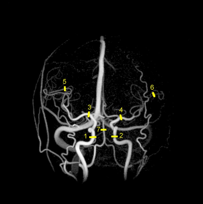
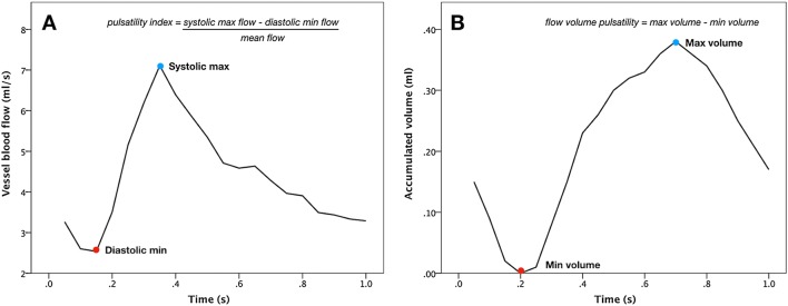
Cerebral small vessel disease (SVD) is a major cause of stroke and cognitive impairment. However, the underlying mechanisms behind SVD are still poorly understood. High cerebral arterial pulsatility has been suggested as a possible cause of SVD. In population studies, arterial pulsatility has been linked to white matter hyperintensities (WMH), cerebral atrophy, and cognitive impairment, all features of SVD. In stroke, pulsatility data are scarce and contradictory. The aim of this study was to investigate the relationship between arterial pulsatility and SVD in stroke patients. With a cross-sectional design, 89 patients with acute ischemic stroke or TIA were examined with MRI. A neuropsychological assessment was performed 1 year later. Using 4D flow MRI, pulsatile indices (PI) were calculated for the internal carotid artery (ICA) and middle cerebral artery (M1, M3). Flow volume pulsatility (FVP), a measure corresponding to the cyclic expansion of the arterial tree, was calculated for the same locations. These parameters were assessed for associations with WMH volume, brain volume and cognitive function. ICA-FVP was associated with WMH volume (β = 1.67, 95% CI: [0.1, 3.24], p = 0.037). M1-PI and M1-FVP were associated with decreasing cognitive function (β = - 4.4, 95% CI: [- 7.7, - 1.1], p = 0.009 and β = - 13.15, 95% CI: [- 24.26, - 2.04], p = 0.02 respectively). In summary, this supports an association between arterial pulsatility and SVD in stroke patients, and provides a potential target for further research and preventative treatment. FVP may become a useful biomarker for assessing pulsatile stress with PCMRI and 4D flow MRI.
Keywords: 4D flow MRI; Pulsatile index; Pulsatility; Small vessel disease; White matter hyperintensities.
Conflict of interest statement
The authors declare that there is no conflict of interest.
Figures
References
MeSH terms
Grants and funding
LinkOut - more resources
Full Text Sources
Medical
Research Materials
Miscellaneous
