

Alterations of perineuronal nets in the dorsolateral prefrontal cortex of neuropsychiatric patients
- PMID: 31728775
- PMCID: PMC6856240
- DOI: 10.1186/s40345-019-0161-0
Alterations of perineuronal nets in the dorsolateral prefrontal cortex of neuropsychiatric patients
Abstract
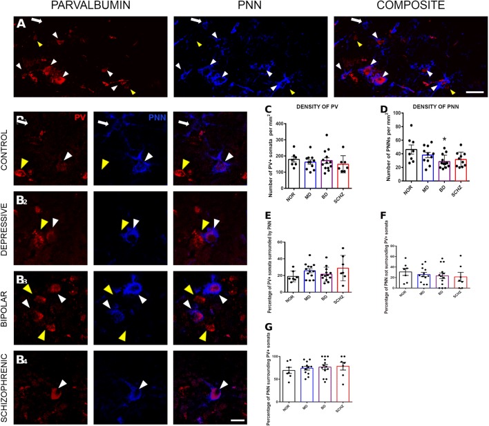
Background: Alterations in the structure and physiology of interneurons in the prefrontal cortex (PFC) are important factors in the etiopathology of different psychiatric disorders. Among the interneuronal subpopulations, parvalbumin (PV) expressing cells appear to be specially affected. Interestingly, during development and adulthood the connectivity of these interneurons is regulated by the presence of perineuronal nets (PNNs), specialized regions of the extracellular matrix, which are frequently surrounding PV expressing neurons. Previous reports have found anomalies in the density of PNNs in the PFC of schizophrenic patients. However, although some studies have described alterations in PNNs in some extracortical regions of bipolar disorder patients, there are no studies focusing on the prefrontocortical PNNs of bipolar or major depression patients. For this reason, we have analyzed the density of PNNs in post-mortem sections of the dorsolateral PFC (DLPFC) from the Stanley Neuropathology Consortium, which includes controls, schizophrenia, bipolar and major depression patients.
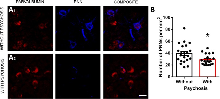
Results: We have not observed differences in the distribution of PV+ cells or PNNs, or in the percentage of PV+ interneurons surrounded by PNNs. The density of PV+ interneurons was similar in all the experimental groups, but there was a significantly lower density of PNNs in the DLPFC of bipolar disorder patients and a tendency towards a decrease in schizophrenic patients. No differences were found when evaluating the density of PV+ cells surrounded by PNNs. Interestingly, when assessing the influence of demographic data, we found an inverse correlation between the density of PNNs and the presence of psychosis.
Conclusions: The present results point to prefrontocortical PNNs and their role in the regulation of neuronal plasticity as putative players in the etiopathology of bipolar disorder and schizophrenia. Our findings also suggest a link between these specialized regions of the extracellular matrix and the presence of psychosis.
Keywords: Bipolar disorder; Major depression; Parvalbumin; Perineuronal nets; Prefrontal cortex; Schizophrenia.
Conflict of interest statement
All authors disclose any actual or potential competing interest including any financial, personal or other relationships with other people or organizations within 3 years of beginning the submitted work that could inappropriately influence, or be perceived to influence, this work.
Figures
References
Grants and funding
LinkOut - more resources
Full Text Sources
Miscellaneous
