Treatment-induced lesions in newly diagnosed glioblastoma patients undergoing chemoradiotherapy and heat-shock protein vaccine therapy
- PMID: 31728884
- PMCID: PMC6939141
- DOI: 10.1007/s11060-019-03336-3
Treatment-induced lesions in newly diagnosed glioblastoma patients undergoing chemoradiotherapy and heat-shock protein vaccine therapy
Abstract
Objectives: Treatment-induced lesions represent a great challenge in neuro-oncology. The aims of this study were (i) to characterize treatment induced lesions in glioblastoma patients treated with chemoradiotherapy and heat-shock protein (HSP) vaccine and (ii) to evaluate the diagnostic accuracy of diffusion weighted imaging for differentiation between treatment-induced lesions and tumor progression.
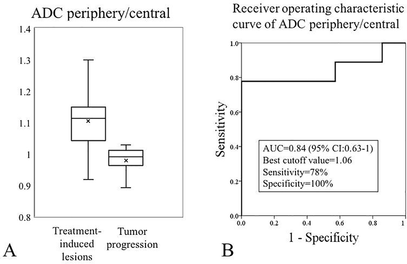
Methods: Twenty-seven patients with newly diagnosed glioblastoma treated with HSP vaccine and chemoradiotherapy were included. Serial magnetic resonance imaging evaluation was performed to detect treatment-induced lesions and assess their growth. Quantitative analysis of the apparent diffusion coefficient (ADC) was performed to discriminate treatment-induced lesions from tumor progression. Mann-Whitney U-test and receiver operating characteristic (ROC) curves were used for analysis.
Results: Thirty-three percent of patients developed treatment-induced lesions. Five treatment-related lesions appeared between end of radiotherapy and the first vaccine administration; 4 lesions within the first 4 months from vaccine initiation and 1 at 3.5 years. Three patients with pathology proven treatment-induced lesions showed a biphasic growth pattern progressed shortly after. ADC ratio between the peripheral enhancing rim and central necrosis showed an accuracy of 0.84 (95% CI 0.63-1) for differentiation between progression and treatment-induced lesions.
Conclusion: Our findings do not support the iRANO recommendation of a 6-month time window in which progressive disease should not be declared after immunotherapy initiation. A biphasic growth pattern of pathologically proven treatment-induced lesions was associated with a dismal prognosis. The presence of lower ADC values in the central necrotic portion of the lesions compared to the enhancing rim shows high specificity for detection of treatment-induced lesions.
Keywords: Chemoradiotherapy; Glioblastoma; Heat-shock proteins; Immunotherapy; Magnetic resonance imaging.
Conflict of interest statement
Compliance with Ethical Standards:
Conflict of Interest:
Author Paula Alcaide Leon declares that she has no conflict of interest.
Author Marisa Lafontaine declares that she has no conflict of interest.
Author Janine M Lupo declares that she has no conflict of interest.
Author Hideho Okada declares that he has no conflict of interest.
Author Jennifer L Clark declares that she has no conflict of interest.
Author Javier E Villanueva-Meyer declares that he has no conflict of interest.
Figures