. 2020 Jan 13;26(3):729-734.
doi: 10.1002/chem.201904363. Epub 2019 Dec 13.

Simultaneous Production of Psilocybin and a Cocktail of β-Carboline Monoamine Oxidase Inhibitors in "Magic" Mushrooms

Affiliations

Simultaneous Production of Psilocybin and a Cocktail of β-Carboline Monoamine Oxidase Inhibitors in "Magic" Mushrooms

Felix Blei et al. Chemistry. .

Abstract

The psychotropic effects of Psilocybe "magic" mushrooms are caused by the l-tryptophan-derived alkaloid psilocybin. Despite their significance, the secondary metabolome of these fungi is poorly understood in general. Our analysis of four Psilocybe species identified harmane, harmine, and a range of other l-tryptophan-derived β-carbolines as their natural products, which was confirmed by 1D and 2D NMR spectroscopy. Stable-isotope labeling with 13 C11 -l-tryptophan verified the β-carbolines as biosynthetic products of these fungi. In addition, MALDI-MS imaging showed that β-carbolines accumulate toward the hyphal apices. As potent inhibitors of monoamine oxidases, β-carbolines are neuroactive compounds and interfere with psilocybin degradation. Therefore, our findings represent an unprecedented scenario of natural product pathways that diverge from the same building block and produce dissimilar compounds, yet contribute directly or indirectly to the same pharmacological effects.

Keywords: alkaloids; ayahuasca; beta-carboline; natural products; psilocybin.


Conflict of interest statement

The authors declare no conflict of interest.

Figures
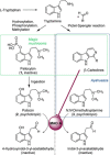
Scheme 1
Scheme 1
Schematic overview on psychoactive principles of Psilocybe mushrooms (green) and ayahuasca (blue), their biosynthetic origin, and their inactivation in the human body by monoamine oxidase (MAO) A.
Figure 1
Figure 1
A) Chromatography of methanolic P. mexicana extracts. Top trace: overlaid extracted ion chromatogram (mass tolerance=0.1 ppm) for the masses of norbaeocystin (m/z=257.0680 [M+H]+, t R=1.33 min), baeocystin (m/z=271.0836 [M+H]+, t R=1.43 min), psilocybin (1, m/z=285.0992 [M+H]+, t R=1.53 min), and psilocin (2, m/z=205.1333 [M+H]+, t R=3.01 min). Below, extracted ion chromatograms for the masses of harmane (4, m/z=183.0916 [M+H]+) and harmine (5, m/z=213.1022 [M+H]+). Bottom: UV/Vis chromatogram (recorded at λ=300 nm, portion from 4.25–5.25 min expanded) and mass spectra. B) HPLC analysis with fluorescence detection. Upper trace: overlaid chromatograms of authentic 4 and 5, lower trace: acidic aqueous P. mexicana mushroom extract. C) HPLC analysis with fluorescence detection. Upper trace: overlaid chromatograms of authentic 47, traces a–d: carpophores of P. cyanescens, P. cubensis FSU12410, P. cubensis FSU12407, and P. semilanceata, respectively. Trace e: P. mexicana sclerotia, traces f and g: P. mexicana and P. cubensis mycelium. D) Chemical structures of β‐carbolines identified as Psilocybe natural products during this study, and of known Psilocybe indole alkaloids baeocystin, norbaeocystin, and norpsilocin.
Figure 2
Figure 2
LC‐MS analysis of P. mexicana mycelial extracts after 13C stable‐isotope labeling. The generic labeling pattern is shown by red carbon atoms. UHPLC chromatograms were recorded at λ=300 nm. Top trace: overlaid chromatograms of standards 410. Center trace: culture grown with unlabeled l‐tryptophan (control). Bottom trace: culture grown in the presence of 13C11l‐tryptophan. Below, HR‐ESI‐MS spectra are shown. Blue: spectra for t R=4.26–4.28 min with coeluting 6 and the isomer of 8 (panel A: unlabeled, panel B: 13C‐labeled situation). Green: spectra for t R=4.50–4.52 min showing 4 and 9/10 coeluting, panel C: unlabeled, panel D: 13C‐labeled. Red: spectra for t R=4.94 min showing 7, panel E: unlabeled, panel F: 13C‐labeled. Upper right: UV/Vis spectra of 4 and collective spectra of the β‐carbolines, detected at t R=4.50 min.
Figure 3
Figure 3
MALDI‐MS imaging of P. cubensis mycelium. The image was taken to detect m/z 183.1(±0.7) Da, i.e., the mass of 4 [M+H]+, and a portion was overlaid on a photograph of the mycelium. Peripheral areas of the mycelium showed highest abundance (red). The image was digitally optimized for brightness which sets the maximum intensity to 60 % of the initial image.

References

    1. Schultes R. E., Amer. Anthropol. 1940, 42, 429–443.
    1. Fricke J., Lenz C., Wick J., Blei F., Hoffmeister D., Chem. Eur. J. 2019, 25, 897–903. - PubMed
    1. None
    1. Hofmann A., Heim R., Brack A., Kobel H., Experientia 1958, 14, 107–109; - PubMed
    1. Hofmann A., Heim R., Brack A., Kobel H., Frey A., Ott H., Petrzilka T., Troxler F., Helv. Chim. Acta 1959, 42, 1557–1572.

MeSH terms