. 2019 Nov 15;10(1):5189.
doi: 10.1038/s41467-019-13209-y.

Cohesin cleavage by separase is enhanced by a substrate motif distinct from the cleavage site

Affiliations

Cohesin cleavage by separase is enhanced by a substrate motif distinct from the cleavage site

Laura E Rosen et al. Nat Commun. .

Abstract

Chromosome segregation begins when the cysteine protease, separase, cleaves the Scc1 subunit of cohesin at the metaphase-to-anaphase transition. Separase is inhibited prior to metaphase by the tightly bound securin protein, which contains a pseudosubstrate motif that blocks the separase active site. To investigate separase substrate specificity and regulation, here we develop a system for producing recombinant, securin-free human separase. Using this enzyme, we identify an LPE motif on the Scc1 substrate that is distinct from the cleavage site and is required for rapid and specific substrate cleavage. Securin also contains a conserved LPE motif, and we provide evidence that this sequence blocks separase engagement of the Scc1 LPE motif. Our results suggest that rapid cohesin cleavage by separase requires a substrate docking interaction outside the active site. This interaction is blocked by securin, providing a second mechanism by which securin inhibits cohesin cleavage.


Conflict of interest statement

The authors declare no competing interests.

Figures

Fig. 1
Fig. 1
Production of active separase. a Cartoon of the securin-separase complex. The vertical dashed line indicates an approximate delineation between the separase N-terminal helical domain (green) and C-terminal protease domain (blue) containing the active site (orange). The C-terminal region of securin (dark purple) binds in an antiparallel fashion along the length of separase, and begins with the pseudosubstrate motif (red) bound in the separase active site. The N-terminal region of securin (light purple) contains the APC/C degrons. b Diagram of the securin-separase fusion construct. Colors correspond to a. Also depicted are the flexible Gly-Ser linker separating securin and separase (light gray) and the regions of human separase predicted to be intrinsically disordered (IDR, dark gray). See Supplementary Fig. 1a for amino acid sequence. c Purified securin-separase fusion protein was analyzed by SDS–PAGE and stained with Coomassie Blue, using molecular weight markers as indicated. d Purified securin-separase (top) and apo (active) separase (bottom) were analyzed by negative-stain EM, and five representative class averages of each preparation are shown. Scale bar: 40 nM. e Securin-separase binding to fluorescein-labeled DNA was evaluated by fluorescence polarization. Two 50 bp dsDNA molecules with the same base composition but different sequence were tested, as well as a 25 bp molecule. Data points indicate means (±SD) of triplicate samples. Source data are provided in the Source Data file. f Separase activation by the ubiquitin-proteasome system, whereby securin is tagged for degradation and removed, can be recapitulated using an N-terminal ClpXP degron and the bacterial protease ClpXP. g Securin-separase fusion protein was incubated with TEV protease, ATP, and/or the ClpXP ATPase as indicated, and separase activity was measured by cleavage of an 35S-labeled Scc1 fragment (residues 142–300) produced by translation in vitro. h Michaelis–Menten analysis was performed with purified, active separase and the peptide DDREIMREGS, which includes cleavage site 1 in Scc1. The peptide sequence was flanked by the MCA fluorophore and DNP quencher, and cleavage was monitored by an increase in fluorescence. Initial velocity was normalized to enzyme concentration. Data points indicate means (±SD) of triplicate samples. Source data are provided in the Source Data file
Fig. 2
Fig. 2
Identification of a separase docking motif on Scc1. a Diagram of the Scc1 sequence, showing the locations of two separase cleavage sites, LPE motif, and boundaries of truncated constructs evaluated in panels b and c. b 35S-labeled Scc1 fragments were incubated with active or inactive separase as indicated, and reaction products were analyzed by SDS-PAGE and Phosphorimaging. c Separase was incubated with an 35S-labeled Scc1 fragment (aa 142–300) in which the indicated residues were changed to alanines. Reaction products were analyzed by SDS–PAGE and Phosphorimaging. The sequence of the relevant region of Scc1 is shown
Fig. 3
Fig. 3
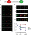
The LPE motif is important for cleavage of a separase biosensor in vivo. a Schematic of the separase biosensor used to evaluate cleavage in vivo, which includes histone H2B, red fluorescent protein (RFP), the indicated Scc1 fragment, and green fluorescent protein (GFP). b Time course of wild-type (WT) biosensor cleavage by separase, showing green fluorescence (left), red fluorescence (center), and merged images (right). Time zero is the last time point before the onset of chromosome segregation. Biosensor cleavage is indicated by reduced green fluorescence relative to red fluorescence. Scale bar: 20 μM. c Representative images showing late anaphase fluorescence of biosensor variants carrying mutations in Scc1 (WT, wild-type, copied from a; NC, non-cleavable mutations at sites 1 and 2; LP → AA, mutations of 255LP; ∆10aa, deletion of aa 251 to 260, which contain the LPE motif). Scale bars: 20 μM. d Quantification of the loss of GFP fluorescence in the four biosensor variants shown in c. Data points indicate means (±SD) from between 15 and 30 cells. Source data are provided in the Source Data file
Fig. 4
Fig. 4
Securin inhibits binding to the LPE motif of Scc1. a Cartoon of the securin-separase fusion protein containing the full separase-binding region of securin (left) or with securin truncated on the C-terminal side of the pseudosubstrate motif (right). The separase active site (orange) and pseudosubstrate motif (red) are indicated. b Diagram of the human securin sequence, indicating the locations of the pseudosubstrate sequence (EIEKFFP), all LP sites including the 130LPE motif, and the positions of the three truncations tested. See Supplementary Figs. 1b and 7 for amino acid sequences. c Michaelis–Menten analysis was performed with the three indicated securinΔ-separase fusion proteins and compared with purified separase lacking securin. Initial velocity was normalized by enzyme concentration. Data points indicate means (±SD) of triplicate samples. See Supplementary Fig. 8 for a control experiment with securin that includes the pseudosubstrate motif. Source data are provided in the Source Data file. d 35S-labeled Scc1 fragments (aa 142–300), with or without mutations in the 255LPE motif, were incubated with the three indicated securinΔ-separase fusion proteins or with purified separase lacking securin. Reaction products were analyzed by SDS-PAGE and Phosphorimaging. e The pseudosubstrate motif in securin was converted to a separase cleavage site using two point mutations (118FP to RE). Separase was incubated with an 35S-labeled securinRE fragment (aa 93–150) containing these mutations as well as mutations in the indicated LP motifs. Reaction products were analyzed by SDS–PAGE and Phosphorimaging. Uncropped autoradiograph is provided in the Source Data file. f Sequence alignment of securin pseudosubstrate motifs (red), indicating the downstream conserved LPE motifs (gray). g Cartoon of the securin-separase complex, illustrating the pseudosubstrate motif interaction with the active site and the LPE motif interaction with the separase exosite

References

    1. Uhlmann F, Lottspeich F, Nasmyth K. Sister-chromatid separation at anaphase onset is promoted by cleavage of the cohesin subunit Scc1. Nature. 1999;400:37–42. doi: 10.1038/21831. - DOI - PubMed
    1. Uhlmann F, Wernic D, Poupart MA, Koonin EV, Nasmyth K. Cleavage of cohesin by the CD clan protease separin triggers anaphase in yeast. Cell. 2000;103:375–386. doi: 10.1016/S0092-8674(00)00130-6. - DOI - PubMed
    1. Nasmyth K, Haering CH. Cohesin: its roles and mechanisms. Annu Rev. Genet. 2009;43:525–558. doi: 10.1146/annurev-genet-102108-134233. - DOI - PubMed
    1. Lin Z, Luo X, Yu H. Structural basis of cohesin cleavage by separase. Nature. 2016;532:131–134. doi: 10.1038/nature17402. - DOI - PMC - PubMed
    1. Luo S, Tong L. Molecular mechanism for the regulation of yeast separase by securin. Nature. 2017;542:255–259. doi: 10.1038/nature21061. - DOI - PMC - PubMed

Publication types