. 2019 Nov 15;10(1):5204.
doi: 10.1038/s41467-019-13205-2.

Virus-borne mini-CRISPR arrays are involved in interviral conflicts

Affiliations

Virus-borne mini-CRISPR arrays are involved in interviral conflicts

Sofia Medvedeva et al. Nat Commun. .

Abstract

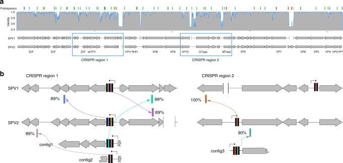
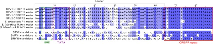
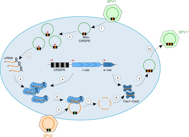
CRISPR-Cas immunity is at the forefront of antivirus defense in bacteria and archaea and specifically targets viruses carrying protospacers matching the spacers catalogued in the CRISPR arrays. Here, we perform deep sequencing of the CRISPRome-all spacers contained in a microbiome-associated with hyperthermophilic archaea of the order Sulfolobales recovered directly from an environmental sample and from enrichment cultures established in the laboratory. The 25 million CRISPR spacers sequenced from a single sampling site dwarf the diversity of spacers from all available Sulfolobales isolates and display complex temporal dynamics. Comparison of closely related virus strains shows that CRISPR targeting drives virus genome evolution. Furthermore, we show that some archaeal viruses carry mini-CRISPR arrays with 1-2 spacers and preceded by leader sequences but devoid of cas genes. Closely related viruses present in the same population carry spacers against each other. Targeting by these virus-borne spacers represents a distinct mechanism of heterotypic superinfection exclusion and appears to promote archaeal virus speciation.


Conflict of interest statement

The authors declare no competing interests.

Figures

Fig. 1
Fig. 1
Characteristics of the analyzed spacer collections. a The four principal repeat sequences found in Sulfolobales; the color-coded repeat sequences and the corresponding PAMs are shown. The last eight nucleotides shared between CRISPR-A and CRISPR-D are underlined. IUPAC nucleotide code is used to show variations in nucleotide sequence of CRISPR repeats in different Sulfolobales genomes: H = A, C or T; Y = C or T; R = A or G; M = A or C; W = A or T; K = G or T. b Intersection of the Beppu spacer collection and spacers from sequenced Sulfolobales isolates available in public databases. The numbers represent the actual number of spacer clusters. c A circular diagram of spacers amplified from the Beppu hot spring and spacers from the Sulfolobales isolates clustered by the country of origin. Spacers belonging to arrays with the four principal repeat sequences found in Sulfolobales are indicated by identical colors matching those used in (a). Spacers that differ from each other by less than two nucleotides are connected by lines whose colors correspond to colors indicating CRISPR consensuses. Black lines connect spacers shared by arrays of different types. The outer gray histograms represent the abundance of CRISPR spacers in log10 scale. YNP, Yellowstone National Park, United States. d The bar plot showing the numbers of protospacers found in Sulfolobales host genomes, plasmids and viruses. The stars indicate values that differ significantly (chi square test, P-value < 0.001) from the expectation. Colors represent spacers associated with different CRISPR consensuses, as in (a). e The bar plot shows the numbers of protospacers found in the top 10 targeted Sulfolobales viruses. Names of viruses isolated in Beppu, Japan are highlighted with violet color. Source data for c, d, and e are provided as a Source Data file
Fig. 2
Fig. 2
The violin plots show the density of the distribution of spacer abundances in the environmental sample and enrichment cultures established from samples J14 and J15. In the J14 sample, the enrichment culture established in the Acidianus-favoring medium is separated from those established in the Sulfolobus/Saccharolobus-favoring medium by a dashed line. Plots in each row represent spacers, associated with different CRISPR consensuses and color-coded as in Fig. 1a. Plots in each row are scaled to have the same area. Log10 scale for the abundance values was used. Source data are provided as a Source Data file
Fig. 3
Fig. 3
Mini-CRISPR arrays in SPV1 and SPV2 genomes. a Comparison of the SPV1 and SPV2 genomes. Genes are represented with arrows following the direction of transcription. Deletions in one of the two genomes with respect to the other are indicated as gaps bordered with vertical lines. Gray histogram above the genome maps shows the identity calculated in 50 bp window from the SPV1-SPV2 nucleotide alignment. Locations of protospacers are showed as colored bars at the top of the figure. The regions zoomed-in in (b) are boxed. ZnF, zinc finger protein; (w)HTH, (winged) helix-turn-helix domain-containing protein; RHH, ribbon helix helix domain-containing protein; MTase, methyltransferase; GTase, glycosyltransferase; VP, virion protein. Source data are provided as a Source Data file. b Zoom-in on two regions of the SPV1 and SPV2 genomes carrying mini-CRISPR arrays (CRISPR region 1 and CRISPR region 2). Black bars represent CRISPR repeat. The predicted promoters in the leader sequences are indicated with broken arrows. Positions of hits of spacers from mini-CRISPR arrays carried by SPV-like viruses are shown with colored bars and arrows link the spacers and the corresponding protospacers. Identities between spacer and protospacers are indicated next to the protospacer bars. Three mini-CRISPR arrays found in the virome data are shown below the corresponding regions of alignment and labeled as contigs 1 to 3
Fig. 4
Fig. 4
Alignment of the loci including the leader sequences and CRISPR repeats associated with the Saccharolobus CRISPR arrays and virus-borne mini-CRISPR arrays. The bottom 3 sequences correspond to stand-alone CRISPR repeats. BRE and TATA elements found in the promoters of the leader sequences are boxed
Fig. 5
Fig. 5
Mini-CRISPR arrays in viral genomes. a mini-CRISPR arrays predicted from the CRISPRome data (labeled as a1 through a15). Identity of spacers to SPV1 or SPV2 genomes is color-coded with the scale provided at the bottom of the figure. Mini-CRISPR arrays found in SPV1, SPV2 and 3 viral contigs shown in Fig. 3b are labeled on the left. Pink and blue boxes show two pairs of identical spacers. b Mini-CRISPR arrays and standalone repeats in Sulfolobales viruses and plasmids. c Total abundance of SPV1 and SPV2 matching spacers from long (cellular) CRISPR arrays and mini-CRISPR (viral) arrays from SPV1 and SPV2 as well as minor SPV strains. Source data are provided as a Source Data file
Fig. 6
Fig. 6
Proposed mechanism of CRISPR-mediated interviral conflict. 1: SPV1 carrying mini-CRISPR array with a spacer against SPV2 infects a susceptible host; 2: SPV1 genome replication results in increased copy number of the mini-CRISPR arrays compared to the chromosomal CRISPR arrays; 3: transcription of the mini-CRISPR arrays; 4: viral crRNAs are complexed with the cellular interference module (i-cas); 5: secondary coinfection of the same cell with SPV2; 6 and 7: SPV2 genome is recognized and degraded by the effector module; 8: fragments of the SPV2 genome are loaded on the cellular Cas1-Cas2 adaptation complex (a-cas); 9: mini-CRISPR array of SPV1 is upgraded with new spacers against SPV2; 10: New variant of SPV1 (SPV1*) is released and spreads the immunity in the population

References

    1. Koonin EV, Krupovic M. The depths of virus exaptation. Curr. Opin. Virol. 2018;31:1–8. doi: 10.1016/j.coviro.2018.07.011. - DOI - PubMed
    1. Forterre P, Prangishvili D. The great billion-year war between ribosome- and capsid-encoding organisms (cells and viruses) as the major source of evolutionary novelties. Ann. N. Y. Acad. Sci. 2009;1178:65–77. doi: 10.1111/j.1749-6632.2009.04993.x. - DOI - PubMed
    1. Koonin EV, Dolja VV. A virocentric perspective on the evolution of life. Curr. Opin. Virol. 2013;3:546–557. doi: 10.1016/j.coviro.2013.06.008. - DOI - PMC - PubMed
    1. Breitbart M, Bonnain C, Malki K, Sawaya NA. Phage puppet masters of the marine microbial realm. Nat. Microbiol. 2018;3:754–766. doi: 10.1038/s41564-018-0166-y. - DOI - PubMed
    1. Chow CE, Suttle CA. Biogeography of viruses in the sea. Annu. Rev. Virol. 2015;2:41–66. doi: 10.1146/annurev-virology-031413-085540. - DOI - PubMed

Publication types

MeSH terms