. 2019 Nov 15;10(1):322.
doi: 10.1186/s13287-019-1435-2.

Intra-pancreatic tissue-derived mesenchymal stromal cells: a promising therapeutic potential with anti-inflammatory and pro-angiogenic profiles

Affiliations

Intra-pancreatic tissue-derived mesenchymal stromal cells: a promising therapeutic potential with anti-inflammatory and pro-angiogenic profiles

Bashar Khiatah et al. Stem Cell Res Ther. .

Abstract

Background: Human pancreata contain many types of cells, such as endocrine islets, acinar, ductal, fat, and mesenchymal stromal cells (MSCs). MSCs are important and shown to have a promising therapeutic potential to treat various disease conditions.

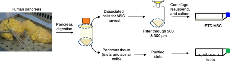
Methods: We investigated intra-pancreatic tissue-derived (IPTD) MSCs isolated from tissue fractions that are routinely discarded during pancreatic islet isolation of human cadaveric donors. Furthermore, whether pro-angiogenic and anti-inflammatory properties of these cells could be enhanced was investigated.

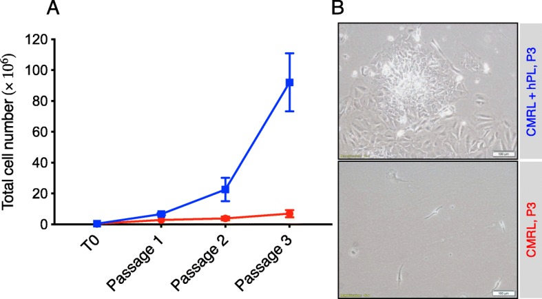
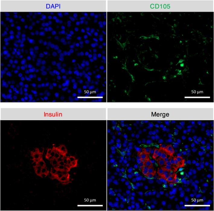
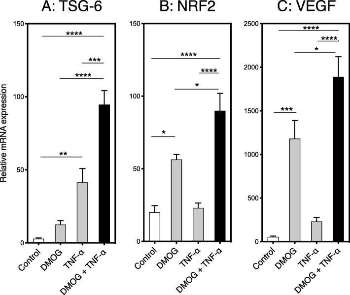
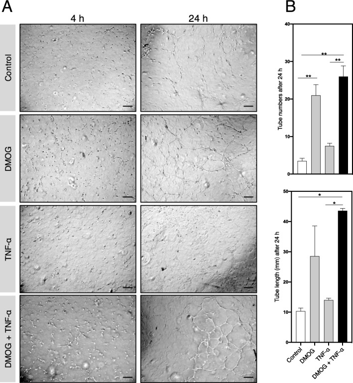
Results: IPTD-MSCs were expanded in GMP-compatible CMRL-1066 medium supplemented with 5% human platelet lysate (hPL). IPTD-MSCs were found to be highly pure, with > 95% positive for CD90, CD105, and CD73, and negative for CD45, CD34, CD14, and HLA-DR. Immunofluorescence staining of pancreas tissue demonstrated the presence of CD105+ cells in the vicinity of islets. IPTD-MSCs were capable of differentiation into adipocytes, chondrocytes, and osteoblasts in vitro, underscoring their multipotent features. When these cells were cultured in the presence of a low dose of TNF-α, gene expression of tumor necrosis factor alpha-stimulated gene-6 (TSG-6) was significantly increased, compared to control. In contrast, treating cells with dimethyloxallyl glycine (DMOG) (a prolyl 4-hydroxylase inhibitor) enhanced mRNA levels of nuclear factor erythroid 2-related factor 2 (NRF2) and vascular endothelial growth factor (VEGF). Interestingly, a combination of TNF-α and DMOG stimulated the optimal expression of all three genes in IPTD-MSCs. Conditioned medium of IPTD-MSCs treated with a combination of DMOG and TNF-α contained higher levels of pro-angiogenic (VEGF, IL-6, and IL-8) compared to controls, promoting angiogenesis of human endothelial cells in vitro. In contrast, levels of MCP-1, a pro-inflammatory cytokine, were reduced in the conditioned medium of IPTD-MSCs treated with a combination of DMOG and TNF-α.

Conclusions: The results demonstrate that IPTD-MSCs reside within the pancreas and can be separated as part of a standard islet-isolation protocol. These IPTD-MSCs can be expanded and potentiated ex vivo to enhance their anti-inflammatory and pro-angiogenic profiles. The fact that IPTD-MSCs are generated in a GMP-compatible procedure implicates a direct clinical application.

Keywords: Angiogenesis; Anti-inflammatory; Mesenchymal stromal cells; NRF2; TSG-6; Type 1 diabetes; VEGF.


Conflict of interest statement

BK, MQ, HTK, and IHA have patents pending related to this study. The other authors declare that they have no competing interests.

Figures

Fig. 1
Fig. 1
A schematic diagram showing the steps for isolating in a cGMP facility intra-pancreatic tissue-derived cells during human islet isolation
Fig. 2
Fig. 2
IPTD cells resemble MSCs in culture and can be cryopreserved Phase contrast microscopy of IPTD-MSCs cells cultured in CMRL-1066 medium supplemented with 5% hPL. a Passage 3 cell culture on day 3; b passage 5 cell culture on day 3; c passage 3 cells after 9 months of cryopreservation, thawing, and culture on day 3. Representative images are presented. Scale bar, 200 μm
Fig. 3
Fig. 3
The growth of IPTD-MSCs is enhanced by a culture medium supplemented with hPL. a Total cell numbers during expansion under the designated culture conditions. hPL was essential for the expansion of cells in vitro. The results shown were from three different tissue donors; b A representative photomicrograph of passage 3 cells cultured in CMRL-1066 culture medium with (green) and without (red) 5% hPL. Scale bar, 100 μm
Fig. 4
Fig. 4
IPTD-MSCs display a cell-surface protein profile consistent with classic MSCs. a Flow cytometry analysis of cell-surface protein expression of bone marrow-derived MSCs and IPTD-MSCs. Data are representative of four pancreas donors. b Immunofluorescent staining for CD105 protein expression (green) in paraffin sections of IPTD-MSCs (passages 3 and 5) grown in the presence of hPL. Scale bar = 50 μm. DAPI stains nuclei
Fig. 5
Fig. 5
Cells expressing CD105 are found in non-digested pancreatic tissue. Double immunofluorescent staining for CD105 (green) and insulin (red) revealed that CD105-positive cells are present in the pancreatic tissue and located adjacent to the insulin-expressing islets. Photomicrographs were obtained using a Z1 microscope (Carl Zeiss) at flourescence wavelengths of 488 nm (CD 105) and 647 nm (insulin). Images are representative of 3 separate experiments. Scale bars, 50 μm
Fig. 6
Fig. 6
IPTD-MSCs can be differentiated into adipogenic, chondrogenic, and osteogenic cells. (a) Passage 3 of IPTD-MSCs prior to differentiation. (b) Lipid droplets are detected after staining with Oil Red O indicating that IPTD-MSCs are undergoing adipogenic differentiation 21 days post culture. (c) The presence of cartilage is confirmed after staining with Alcian Blue showing in dark blue color. (d) Calcium deposition is detected in paraffin-fixed tissue section of the cells cultured in chondrogenic media after staining with Alcian Blue. (e) Calcium deposition is detected in paraffin-fixed tissue section of the cells cultured in osteogenic medium after staining with von Kossa
Fig. 7
Fig. 7
IPTD-MSCs treated with TNF-α and DMOG display increased mRNA levels of anti-inflammatory and pro-angiogenic genes. a TSG-6, b NRF2, and c VEGF mRNA levels from IPTD-MSCs treated with TNF-α (10 ng/mL) and/or DMOG (1 mM). Three independent donors were tested. The data are expressed as mean ± SEM. *p < 0.05; **p < 0.01; ***p < 0.001; ****p < 0.0001
Fig. 8
Fig. 8
IPTD-MSCs treated with TNF-α and DMOG produce increased levels of several cytokines. Relative changes of the indicated cytokines and growth factors found in medium from conditioned and control IPTD-MSCs. Y-axis is logarithmic. Four independent experiments were performed. The data are expressed as mean ± SEM. *p < 0.05; **p < 0.01; ***p < 0.001; ****p < 0.0001; n.s. not significant
Fig. 9
Fig. 9
IPTD-MSC-derived media stimulates endothelial tube formation. a Representative photographs of endothelial cells cultured and treated with medium from conditioned and control IPTD-MSCs at 4 and 24 h post-culture. Scale bar, 500 μm. b Quantification is presented as total tube number and total tube length after 24 h. Duplicate samples were performed. Representative microscopic images are presented

Similar articles

Cited by

References

    1. Roberts I. Mesenchymal stem cells. Vox Sang. 2004;87(Suppl 2):38–41. doi: 10.1111/j.1741-6892.2004.00452.x. - DOI - PubMed
    1. Olsen TR, Ng KS, Lock LT, et al. Peak MSC-are we there yet? Front Med (Lausanne) 2018;5:178. doi: 10.3389/fmed.2018.00178. - DOI - PMC - PubMed
    1. Abdi R, Fiorina P, Adra CN, et al. Immunomodulation by mesenchymal stem cells: a potential therapeutic strategy for type 1 diabetes. Diabetes. 2008;57:1759–1767. doi: 10.2337/db08-0180. - DOI - PMC - PubMed
    1. Guan LX, Guan H, Li HB, et al. Therapeutic efficacy of umbilical cord-derived mesenchymal stem cells in patients with type 2 diabetes. Exp Ther Med. 2015;9:1623–1630. doi: 10.3892/etm.2015.2339. - DOI - PMC - PubMed
    1. Liu X, Zheng P, Wang X, et al. A preliminary evaluation of efficacy and safety of Wharton's jelly mesenchymal stem cell transplantation in patients with type 2 diabetes mellitus. Stem Cell Res Ther. 2014;5:57. doi: 10.1186/scrt446. - DOI - PMC - PubMed

Publication types

MeSH terms