. 2020 Jan 1:101:237-248.
doi: 10.1016/j.actbio.2019.11.021. Epub 2019 Nov 13.

Biomaterial-mediated reprogramming of monocytes via microparticle phagocytosis for sustained modulation of macrophage phenotype

Affiliations

Biomaterial-mediated reprogramming of monocytes via microparticle phagocytosis for sustained modulation of macrophage phenotype

Kathryn L Wofford et al. Acta Biomater. .

Abstract

Monocyte-derived macrophages orchestrate tissue regeneration by homing to sites of injury, phagocytosing pathological debris, and stimulating other cell types to repair the tissue. Accordingly, monocytes have been investigated as a translational and potent source for cell therapy, but their utility has been hampered by their rapid acquisition of a pro-inflammatory phenotype in response to the inflammatory injury microenvironment. To overcome this problem, we designed a cell therapy strategy where monocytes are exogenously reprogrammed by intracellularly loading the cells with biodegradable microparticles containing an anti-inflammatory drug in order to modulate and maintain an anti-inflammatory phenotype over time. To test this concept, poly(lactic-co-glycolic) acid microparticles were loaded with the anti-inflammatory drug dexamethasone (Dex) and administered to primary human monocytes for four hours to facilitate phagocytic uptake. After removal of non-phagocytosed microparticles, microparticle-loaded monocytes differentiated into macrophages and stored the microparticles intracellularly for several weeks in vitro, releasing drug into the extracellular environment over time. Cells loaded with intracellular Dex microparticles showed decreased expression and secretion of inflammatory factors even in the presence of pro-inflammatory stimuli up to 7 days after microparticle uptake compared to untreated cells or cells loaded with blank microparticles, without interfering with phagocytosis of tissue debris. This study represents a new strategy for long-term maintenance of anti-inflammatory macrophage phenotype using a translational monocyte-based cell therapy strategy without the use of genetic modification. Because of the ubiquitous nature of monocyte-derived macrophage involvement in pathology and regeneration, this strategy holds potential as a treatment for a vast number of diseases and disorders. STATEMENT OF SIGNIFICANCE: We report a unique and translational strategy to overcome the challenges associated with monocyte- and macrophage-based cell therapies, in which the cells rapidly take on inflammatory phenotypes when administered to sites of injury. By intracellularly loading monocytes with drug-loaded microparticles prior to administration via phagocytosis, we were able to inhibit inflammation while preserving functional behaviors of human primary macrophages derived from those monocytes up to seven days later. To our knowledge, this study represents the first report of reprogramming macrophages to an anti-inflammatory phenotype without the use of genetic modification.

Keywords: Cell-microparticle interactions; Dexamethasone; Inflammation; Intracellular microparticles; Monocyte-derived macrophages; Phagocytosis.


Figures

Figure 1.
Figure 1.. Biomaterial-mediated monocyte cell therapy strategy.
(A) Monocytes are isolated from the patient’s blood and incubated with immunomodulatory microparticles, which are rapidly phagocytosed. (B) Microparticle-loaded cells can be administered systemically or locally to the site of injury for a minimally-invasive, autologous treatment. (C) Degradation of the immunomodulatory microparticles over time allows intracellular release of the drug, dexamethasone (Dex), where it can modulate macrophage phenotype.
Figure 2.
Figure 2.. Phagocytosed microparticles release model drugs intracellularly for several weeks.
(A) Size distribution of single-emulsion PLGA microparticles fabricated with no drug (Blank), with Dex, or with the fluorescent model drug tetramethylrhodamine (TRITC). (B) Cells loaded with fluorescent microparticles can be imaged over time. (C) Cell area, and thus monocyte-to-macrophage differentiation, is not affected by intracellular microparticle loading. Box and whisker plot represents all data ranging from the minimum to the maximum. (D-E) Intracellular fluorescent microparticles were quantified on a single cell level for number of intracellular microparticles and intracellular microparticle intensity over time per cell (1589 cells analyzed from n=8 experimental replicates). (F-J) Five days after TRITC microparticle administration, cells were stained for nuclei (DAPI, blue) and BuGR2, a glucocorticoid receptor (green) that can be found in the cytoplasm, for imaging along with TRITC (red; n=3). Areas where TRITC signal co-localized with the BuGR2 signal are represented in white. (K) Conditioned media from macrophages loaded with fluorescent microparticles was quantified spectrophotometrically to assess the concentration of extracellular TRITC release from the cells over time (n=22). Scale bars = 50 μm. (L-M) Representative images of untreated macrophages or TRITC microparticle-loaded macrophage morphology and density after 43 days of in vitro culture. Data represent mean ± SD for all graphs. Box and whisker plots represent 5–95th percentile of the data with the remaining data plotted as points. Scale bars = 50 μm.
Figure 3.
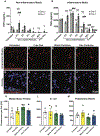
Figure 3.. Intracellular Dex microparticles modulate cell behavior one week after microparticle treatment.
(A) Release profile of Dex from microparticles in PBS (n=5). (B) Dex content in the cell culture media (n=3 in blank group and n=6 in treatment groups) following Dex microparticle administration to cells. Media was changed on days 3, 5, and 7. (C-J) Representative images of cells stained for CD163 and CCR7 or stained for MerTk and CCR2. (K-N) Image quantification of staining for the surface receptors CD163, CCR7, MerTk, and CCR2 (n=4). Data represent mean ± SEM for all graphs. Statistical analyses were completed by applying one-way ANOVA with Tukey’s post hoc test. *denotes p<0.05 and ** denotes p<0.01. Scale bar = 50 μm.
Figure 4.
Figure 4.. Intracellular Dex microparticles modulate cell behavior even in the presence of inflammatory stimuli.
Tumor necrosis factor alpha (TNFα) protein secretion was measured in conditioned media from microparticle-treated cells in (A) non-inflammatory or (B) inflammatory media (n=6). Data represent mean ± SEM. Two-way ANOVAs with Tukey’s post hoc tests were completed using p values corrected for multiple comparisons. *denotes p<0.05, **denotes p<0.01, ***denotes p<0.001, denotes significant differences relative to untreated controls on day 5, and #denotes significant differences relative to untreated controls on day 7. (C-J) Representative images of DiD-labeled monocytes that were untreated, treated with continuous free Dex, treated with blank microparticles, or treated with Dex microparticles for four hours prior to removal of non-phagocytosed microparticles, and then cultured in inflammatory media conditions for 5 days prior to incubation with phagocytosis targets. Phagocytic uptake of (K) myelin basic protein, (L) E. coli, or (M) polystyrene beads within six hours was quantified across treatment condition (between 6 and 27 cells analyzed and averaged from each experimental replicate with n=4 experimental replicates). Data represent mean ± SEM for all graphs. Statistical analyses were completed by applying one-way ANOVA with Tukey’s post hoc analysis with corrected p values. No statistical differences were detected. Scale bar = 50 μm.
Figure 5.
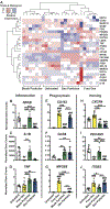
Figure 5.. Intracellular microparticles modulate macrophage gene expression.
(A) Post-processed gene expression was plotted in a heatmap with scaling by rows. Dendrogram organization of the heatmap columns (with each column representing a replicate from a treatment condition) was employed to organize samples according to similar gene expression profiles, clustering columns with similar trends in close proximity to one another (n=6). A subset of genes related to (B-D) inflammation, (E-G) phagocytosis, and (H-J) homing were plotted. All genes analyzed can be found in the supplementary figures 5 and 6. One-way ANOVA statistical analyses were completed on transformed data from each of the data sets. To facilitate interpretation, non-transformed data were plotted in column graphs. Tukey’s post hoc analyses with corrections for multiple comparisons were completed as appropriate. *denotes p<0.05, **denotes p<0.01, ***denotes p<0.001, and #denotes significant differences relative to untreated controls.

References

    1. Wynn TA, Vannella KM, Macrophages in Tissue Repair, Regeneration, and Fibrosis, Immunity. 44 (2016) 450–462. doi:10.1016/j.immuni.2016.02.015. - DOI - PMC - PubMed
    1. Lech M, Anders HJ, Macrophages and fibrosis: How resident and infiltrating mononuclear phagocytes orchestrate all phases of tissue injury and repair, Biochim. Biophys. Acta - Mol. Basis Dis 1832 (2013) 989–997. doi:10.1016/j.bbadis.2012.12.001. - DOI - PubMed
    1. Wattananit S, Tornero D, Graubardt N, Memanishvili T, Monni E, Tatarishvili J, Miskinyte G, Ge R, Ahlenius H, Lindvall O, Schwartz M, Kokaia Z, Monocyte-Derived Macrophages Contribute to Spontaneous Long-Term Functional Recovery after Stroke in Mice, J. Neurosci 36 (2016) 4182–4195. doi:10.1523/JNEUROSCI.4317-15.2016. - DOI - PMC - PubMed
    1. Shechter R, London A, Varol C, Raposo C, Cusimano M, Yovel G, Rolls A, Mack M, Pluchino S, Martino G, Jung S, Schwartz M, Infiltrating blood-derived macrophages are vital cells playing an anti-inflammatory role in recovery from spinal cord injury in mice., PLoS Med. 6 (2009) e1000113. doi:10.1371/journal.pmed.1000113. - DOI - PMC - PubMed
    1. Aderem A, Underhill DM, Mechanisms of Phagocytosis in Macrophages, Annu. Rev. Immunol 17 (1999) 593–623. doi:10.1146/annurev.immunol.17.1.593. - DOI - PubMed

Publication types

MeSH terms

Substances