. 2019 Oct 25;20(21):5309.
doi: 10.3390/ijms20215309.

Comprehensive Analysis of the Chitinase Gene Family in Cucumber (Cucumis sativus L.): From Gene Identification and Evolution to Expression in Response to Fusarium oxysporum

Affiliations

Comprehensive Analysis of the Chitinase Gene Family in Cucumber (Cucumis sativus L.): From Gene Identification and Evolution to Expression in Response to Fusarium oxysporum

Ezra S Bartholomew et al. Int J Mol Sci. .

Abstract

Chitinases, a subgroup of pathogenesis-related proteins, are responsible for catalyzing the hydrolysis of chitin. Accumulating reports indicate that chitinases play a key role in plant defense against chitin-containing pathogens and are therefore good targets for defense response studies. Here, we undertook an integrated bioinformatic and expression analysis of the cucumber chitinases gene family to identify its role in defense against Fusarium oxysporum f. sp. cucumerinum. A total of 28 putative chitinase genes were identified in the cucumber genome and classified into five classes based on their conserved catalytic and binding domains. The expansion of the chitinase gene family was due mainly to tandem duplication events. The expression pattern of chitinase genes was organ-specific and 14 genes were differentially expressed in response to F. oxysporum challenge of fusarium wilt-susceptible and resistant lines. Furthermore, a class I chitinase, CsChi23, was constitutively expressed at high levels in the resistant line and may play a crucial role in building a basal defense and activating a rapid immune response against F. oxysporum. Whole-genome re-sequencing of both lines provided clues for the diverse expression patterns observed. Collectively, these results provide useful genetic resource and offer insights into the role of chitinases in cucumber-F. oxysporum interaction.

Keywords: Cucumis sativus; Fusarium oxysporum; chitinase; gene expression; gene family.


Conflict of interest statement

The authors declare no conflict of interest.

Figures

Figure 1
Figure 1
Phylogenetic and structure analysis of chitinase genes from C. sativus, C. melo and A. thaliana. (A) Phylogenetic tree built using the neighbor-joining (NJ) method in MEGA X. Boot strap values are from 1000 replications. The roman numerals (I–V) representing each chitinase gene class. The numbers at the nodes represent bootstrap percentage values. (B) Schematic representation of exon-intron structure of chitinase genes built using GSDS 2.0. Lengths of exons and introns of each gene are exhibited proportionally. For all genes, black lines represent introns, yellow boxes represent exons and purple boxes represent untranslated regions (UTRs). Intron phases 0, 1 and 2 are represented by blue, green and red triangles, respectively. (C) Schematic representation of conserved motifs of chitinase genes from C. sativus, C. melo and A. thaliana. The conserved motifs in chitinase proteins were identified by MEME software. Grey lines represent the non-conserved sequences, and each motif is indicated by a colored box numbered on the bottom of the figure. The length of motifs in each protein is presented proportionally.
Figure 2
Figure 2
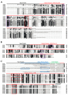
Multiple sequence alignment of the conserved domains of cucumber chitinase. (A) Sequence alignment of GH18 class III chitinases. Shaded amino acids are 50–100% homologous. (B) Sequence alignment of GH18 class V chitinases. Shaded amino acids are 75–100% homologous. (C) Sequence alignment of GH19 (class I, II and IV) chitinases. Shaded amino acids are 75–100% homologous. Amino acid sequences were aligned using DNAMAN software (ver. 6.0). Black and red lines over sequences indicate signal peptides and catalytic domain. While chitin-binding and linker regions are indicated by blue and green line over sequence. Purple boxed residues are conserved. Blue box (dash line) = PS00225, red box = catalytic domain and blue boxes = chitin-binding domain.
Figure 3
Figure 3
Chromosomal location and duplication of chitinase genes in cucumber. The chromosome size is indicated by its relative length using the information from the cucumber 9930 draft genome ver. 2.0 [19]. Black boxes indicated tandem duplication genes. The paralogous genes were written with same colors. Locations are mapped according to base pair start positions. The chromosome number is indicated above each bar and the scale bar on the left is in megabase (Mb).
Figure 4
Figure 4
Heat map representation of chitinase genes expression in different cucumber tissues. The expression data was converted with Log2 (RPKM) to calculate gene expression levels. Gene expression differences are shown in the colors indicated in the scale. The RNA-Seq data used here could be downloaded from http://www.ncbi.nlm.nih.gov/bioproject/PRJNA80169/.
Figure 5
Figure 5
Fusarium wilt infection assay. (A) Disease symptoms of fusarium wilt in cucumber inbred lines 3229 and line 3461. The roots were inoculated with F. oxysporum f. sp. Cucumerinum Owen race 3. Mock plants were treated with distilled water. Red arrows show severe wilting in the stem. (B) Average disease symptom rating of both lines was scored using a disease rating scale (0–4). Values are shown as means ± SD (n = 6). (*, p < 0.05; Student’s t-test).
Figure 6
Figure 6
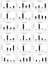
Expression patterns of 18 chitinase genes in C. sativus lines 3229 (susceptible) and 3461 (resistant) after infection with F. oxysporum f. sp. cucumerinum Owen race 3. Data were normalized to the expression level of β-actin. All data points are the means ± SE (n = 3). (*, p < 0.05; Student’s t-test).
Figure 7
Figure 7
Annotation and distribution of single-nucleotide polymorphisms (SNPs) and insertions and deletions (InDels). Distribution of (A) SNPs; and (B) InDels in different genomic regions. (C) Annotation of total large-effect SNPs and InDels in different genic regions. Distribution of (D) SNPs; and (E) InDels in different intergenic and genic regions of chitinase genes from C. sativus lines 3229 vs 3461. The number of synonymous and non-synonymous SNPs detected within the CDS region has also been shown. Distribution of SNPs and InDels were 3229 vs 3461.

References

    1. Renner T., Specht C.D. Molecular Evolution and Functional Evolution of Class I Chitinases for Plant Carnivory in the Caryophyllales. Mol. Biol. Evol. 2012;29:2971–2985. doi: 10.1093/molbev/mss106. - DOI - PubMed
    1. Lombard V., Golaconda Ramulu H., Drula E., Coutinho P.M., Henrissat B. The Carbohydrate-Active Enzymes Database (CAZy) in 2013. Nucleic Acids Res. 2014;42:D490–D495. doi: 10.1093/nar/gkt1178. - DOI - PMC - PubMed
    1. Jain K.K., Kumar A., Shankar A., Pandey D., Chaudhary B., Sharma K.K. De Novo Transcriptome Assembly and Protein Profiling of Copper-Induced Lignocellulolytic Fungus Ganoderma Lucidum MDU-7 Reveals Genes Involved in Lignocellulose Degradation and Terpenoid Biosynthetic Pathways. Genomics. 2019 doi: 10.1016/j.ygeno.2019.01.012. - DOI - PubMed
    1. Tyler L., Bragg J.N., Wu J., Yang X., Tuskan G.A., Vogel J.P. Annotation and Comparative Analysis of the Glycoside Hydrolase Genes in Brachypodium Distachyon. BMC Genom. 2010;11:600. doi: 10.1186/1471-2164-11-600. - DOI - PMC - PubMed
    1. Wang S., Ye X., Chen J., Rao P. A Novel Chitinase Isolated from Vicia Faba and Its Antifungal Activity. Food Res. Int. 2012;45:116–122. doi: 10.1016/j.foodres.2011.10.010. - DOI

MeSH terms

LinkOut - more resources