Review
. 2019 Oct 25;8(11):1782.
doi: 10.3390/jcm8111782.

Induced Pluripotent Stem Cells as Vasculature Forming Entities

Affiliations
Review

Induced Pluripotent Stem Cells as Vasculature Forming Entities

Antonio Palladino et al. J Clin Med. .

Abstract

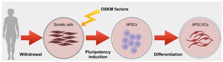
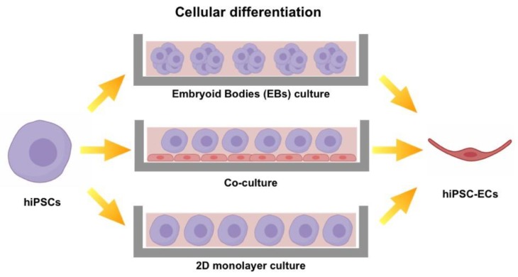
Tissue engineering (TE) pursues the ambitious goal to heal damaged tissues. One of the most successful TE approaches relies on the use of scaffolds specifically designed and fabricated to promote tissue growth. During regeneration the guidance of biological events may be essential to sustain vasculature neoformation inside the engineered scaffold. In this context, one of the most effective strategies includes the incorporation of vasculature forming cells, namely endothelial cells (EC), into engineered constructs. However, the most common EC sources currently available, intended as primary cells, are affected by several limitations that make them inappropriate to personalized medicine. Human induced Pluripotent Stem Cells (hiPSC), since the time of their discovery, represent an unprecedented opportunity for regenerative medicine applications. Unfortunately, human induced Pluripotent Stem Cells-Endothelial Cells (hiPSC-ECs) still display significant safety issues. In this work, we reviewed the most effective protocols to induce pluripotency, to generate cells displaying the endothelial phenotype and to perform an efficient and safe cell selection. We also provide noteworthy examples of both in vitro and in vivo applications of hiPSC-ECs in order to highlight their ability to form functional blood vessels. In conclusion, we propose hiPSC-ECs as the preferred source of endothelial cells currently available in the field of personalized regenerative medicine.

Keywords: angiogenesis; from bench to bedside; induced pluripotent stem cells; tissue engineering; tissue regeneration.


Conflict of interest statement

The authors declare no conflict of interest.

Figures

Figure 1
Figure 1
Sources of endothelial cells (ECs) used in scaffold-based approaches for tissue engineering (TE). HUVECs: Human Umbilical Vein Endothelial Cells, HDMECs: Human Dermal Microvascular Endothelial Cells, ECFCs: Endothelial Colony Forming Cells, ESCs: Embryonic Stem Cells, hIPSC-ECs: Endothelial Cells derived from Human Induced Pluripotent Stem Cells.
Figure 2
Figure 2
Schematic representation of human Induced Pluripotent Stem Cells-EndothelialCells (hiPSC-ECs) generation. Firstly, somatic cells are collected from the patient, then pluripotency is induced by the re-expression of four genes identified by Yamanaka et al. in 2006: OCT-3/4, Sox2, Klf4 and c-Myc (OSKM factors) which are normally inactive in somatic cells. Afterwards, induced Pluripotent Stem Cells (iPSCs) differentiation is induced towards mature Endothelial Cells (ECs).
Figure 3
Figure 3
Diagram representing the different approaches to deliver reprogramming factors OSKM (Oct-3/4, Sox2, Klf4 and c-Myc) to somatic cells. The Integrative approaches: Linear DNA, MuLV (Murine Leukemia Virus), PiggyBac. The Non-Integrative approaches: Episomal Vectors, Adenovirus, SeV (Sendai Virus), Modified RNA and CRISPR-dCas9 Synergistic Activation Mediators (SAM).
Figure 4
Figure 4
Illustration of the main strategies used to differentiate hiPSC into hiPSC-ECs. Embryoid Bodies (EBs): cultured in suspension hiPSCs tend to auto-aggregate in embryoid bodies. Co-culture: hiPSCs are co-cultured with cells able to guide their differentiation into the mature phenotype. Two-dimensional (2D) monolayer culture: hiPSCs are seeded on matrix-coated plates where they are induced to differentiate.

References

    1. Barabaschi G.D.G., Manoharan V. Engineering Mineralized and Load Bearing Tissues. Adv. Exp. Med. Biol. 2015;881:79–94. - PubMed
    1. Baranski J.D., Chaturvedi R.R., Stevens K.R., Eyckmans J., Carvalho B., Solorzano R.D., Yang M.T., Miller J.S., Bhatia S.N., Chen C.S. Geometric control of vascular networks to enhance engineered tissue integration and function. Proc. Natl. Acad. Sci. USA. 2013;110:7586–7591. doi: 10.1073/pnas.1217796110. - DOI - PMC - PubMed
    1. Rossi L., Attanasio C., Vilardi E., De Gregorio M., Netti P.A. Vasculogenic potential evaluation of bottom-up, PCL scaffolds guiding early angiogenesis in tissue regeneration. J. Mater. Sci. Mater. Med. 2016;27:1–11. doi: 10.1007/s10856-016-5720-7. - DOI - PubMed
    1. Natale C.F., Lafaurie-Janvore J., Ventre M., Babataheri A., Barakat A.I. Focal adhesion clustering drives endothelial cell morphology on patterned surfaces. J. R. Soc. Interface. 2019;16:20190263. doi: 10.1098/rsif.2019.0263. - DOI - PMC - PubMed
    1. Fedele C., De Gregorio M., Netti P.A., Cavalli S., Attanasio C. Azopolymer photopatterning for directional control of angiogenesis. Acta Biomater. 2017;63:317–325. doi: 10.1016/j.actbio.2017.09.022. - DOI - PubMed

LinkOut - more resources