Review
. 2019 Nov 15;11(11):612.
doi: 10.3390/pharmaceutics11110612.

The Role of Nanovaccine in Cross-Presentation of Antigen-Presenting Cells for the Activation of CD8+ T Cell Responses

Affiliations
Review

The Role of Nanovaccine in Cross-Presentation of Antigen-Presenting Cells for the Activation of CD8+ T Cell Responses

Cheol Gyun Kim et al. Pharmaceutics. .

Abstract

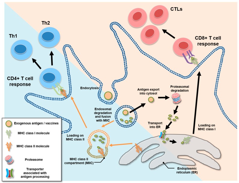
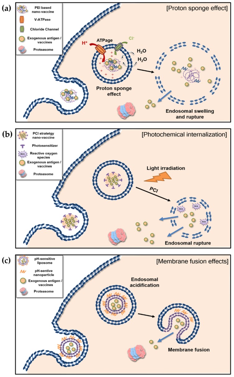
Explosive growth in nanotechnology has merged with vaccine development in the battle against diseases caused by bacterial or viral infections and malignant tumors. Due to physicochemical characteristics including size, viscosity, density and electrostatic properties, nanomaterials have been applied to various vaccination strategies. Nanovaccines, as they are called, have been the subject of many studies, including review papers from a material science point of view, although a mode of action based on a biological and immunological understanding has yet to emerge. In this review, we discuss nanovaccines in terms of CD8+ T cell responses, which are essential for antiviral and anticancer therapies. We focus mainly on the role and mechanism, with particular attention to the functional aspects, of nanovaccines in inducing cross-presentation, an unconventional type of antigen-presentation that activates CD8+ T cells upon administration of exogenous antigens, in dendritic cells followed by activation of antigen-specific CD8+ T cell responses. Two major intracellular mechanisms that nanovaccines harness for cross-presentation are described; one is endosomal swelling and rupture, and the other is membrane fusion. Both processes eventually allow exogenous vaccine antigens to be exported from phagosomes to the cytosol followed by loading on major histocompatibility complex class I, triggering clonal expansion of CD8+ T cells. Advancement of nanotechnology with an enhanced understanding of how nanovaccines work will contribute to the design of more effective and safer nanovaccines.

Keywords: CD8+ T cell; cancer vaccine; cross-presentation; cytotoxic T lymphocyte; dendritic cell; nanovaccine.


Conflict of interest statement

The authors declare no conflicts of interest.

Figures

Figure 1
Figure 1
Schematic diagram for the presentation of an exogenous antigen in DCs. The exogenous antigen, in a general process, is presented by an MHC class II loading pathway (cyan color background). After endocytosis, the endosome-containing antigen goes through the process of maturation, including acidification. Then, MHC class II-containing intracellular vesicles called the MHC class II compartment fuses with the endosome. Through this process, the antigen degraded by proteolytic enzymes and loaded onto MHC class II leads to the presentation to CD4+ T cells. Alternatively, an exogenous antigen could be presented with MHC class I via cross-presentation (yellow background). After endocytosis, the antigen could be exported into cytosol via various pathways (see details in Figure 2). The exported antigens (epitopes) degraded by proteasome are transported to endoplasmic reticulum via transporter associated with antigen processing (TAP). Then, the epitope is loaded onto the pocket of MHC class I resulting in recognition by and activation of CD8+ T cells.
Figure 2
Figure 2
Schematic representation of the mechanism of a nanovaccine inducing cytosolic exportation, leading to cross-presentation: (a) proton sponge effect, (b) photochemical internalization, (c) membrane fusion effect of pH-sensitive liposome. (a) After cationic PEI-based nanovaccines are taken up, and an unsaturated amino group buffers the influxing proton, supplied by a proton pump, v-ATPase. The proton pump continuously transports the protons into endosomes, which results in retention of chloride ions and water. Eventually, osmotic swelling and rupture of endosomes expose the antigen and vaccine compartment to the cytosol; (b) Photochemical internalization based nanovaccines contain a photosensitizer responding to a specific wavelength of light. After light irradiation, the photosensitizer initiates of reactive oxygen species, leading to rupturing of endosomes and cytosolic exports; (c) After endocytosis of liposomes combine with pH-sensitive nanoparticles, early endosome become acidic along with endosomal maturation. The acidic condition destabilizes the pH-sensitive liposomes, leading to enhanced membrane fusion with export of antigens into cytosol.

References

    1. Oyewumi M.O., Kumar A., Cui Z. Nano-microparticles as immune adjuvants: Correlating particle sizes and the resultant immune responses. Expert Rev. Vaccines. 2010;9:1095–1107. doi: 10.1586/erv.10.89. - DOI - PMC - PubMed
    1. Roopngam P.E. Liposome and polymer-based nanomaterials for vaccine applications. Nanomed. J. 2019;6:1–10.
    1. Nakamura T., Harashima H. Integration of nano drug-delivery system with cancer immunotherapy. Ther. Deliv. 2017;8:987–1001. doi: 10.4155/tde-2017-0071. - DOI - PubMed
    1. Pulendran B., Ahmed R. Immunological mechanisms of vaccination. Nat. Immunol. 2011;12:509–517. doi: 10.1038/ni.2039. - DOI - PMC - PubMed
    1. Joffre O.P., Segura E., Savina A., Amigorena S. Cross-presentation by dendritic cells. Nat. Rev. Immunol. 2012;12:557–569. doi: 10.1038/nri3254. - DOI - PubMed