. 2019 Nov 15;9(11):1623.
doi: 10.3390/nano9111623.

Synergistical Use of Electrostatic and Hydrophobic Interactions for the Synthesis of a New Class of Multifunctional Nanohybrids: Plasmonic Magneto-Liposomes

Affiliations

Synergistical Use of Electrostatic and Hydrophobic Interactions for the Synthesis of a New Class of Multifunctional Nanohybrids: Plasmonic Magneto-Liposomes

Gabriela Fabiola Știufiuc et al. Nanomaterials (Basel). .

Abstract

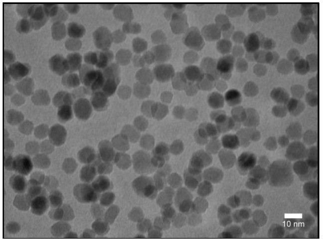
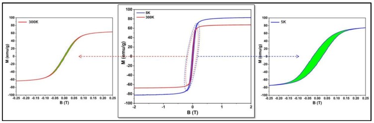
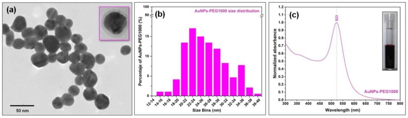
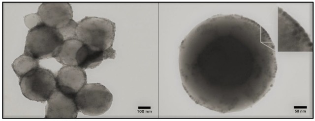
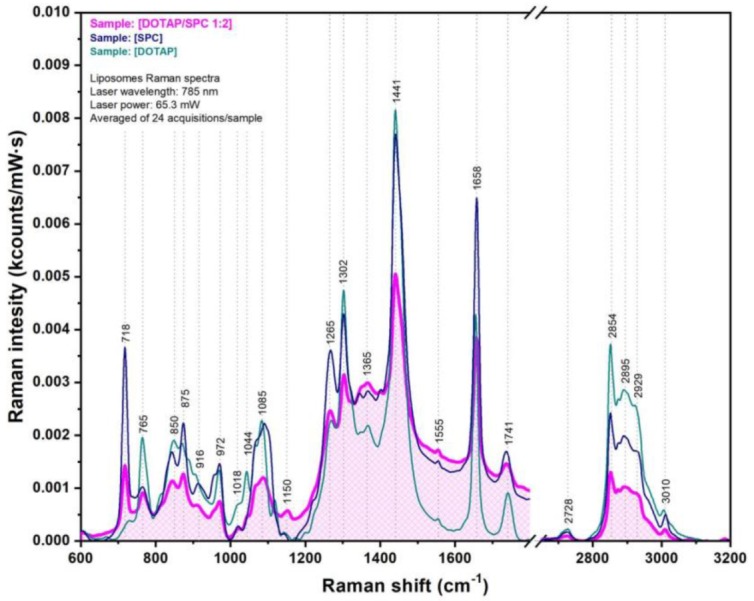
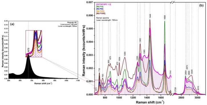
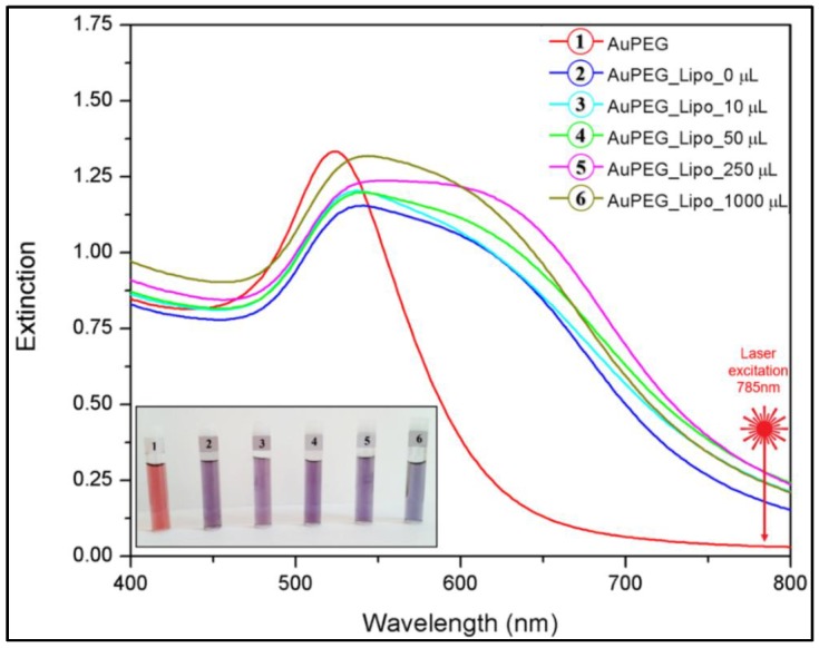
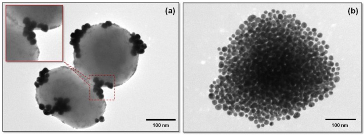
By carefully controlling the electrostatic interactions between cationic liposomes, which already incorporate magnetic nanoparticles in the bilayers, and anionic gold nanoparticles, a new class of versatile multifunctional nanohybrids (plasmonic magneto-liposomes) that could have a major impact in drug delivery and controlled release applications has been synthesized. The experimental results confirmed the successful synthesis of hydrophobic superparamagnetic iron oxide nanoparticles (SPIONs) and polyethylene glycol functionalized (PEGylated) gold nanoparticles (AuNPs). The SPIONs were incorporated in the liposomal lipidic bilayers, thus promoting the formation of cationic magnetoliposomes. Different concentrations of SPIONs were loaded in the membrane. The cationic magnetoliposomes were decorated with anionic PEGylated gold nanoparticles using electrostatic interactions. The successful incorporation of SPIONs together with the modifications they generate in the bilayer were analyzed using Raman spectroscopy. The plasmonic properties of the multifunctional nanohybrids were investigated using UV-Vis absorption and (surface-enhanced) Raman spectroscopy. Their hyperthermic properties were recorded at different frequencies and magnetic field intensities. After the synthesis, the nanosystems were extensively characterized in order to properly evaluate their potential use in drug delivery applications and controlled release as a result of the interaction with an external stimulus, such as an NIR laser or alternating magnetic field.

Keywords: gold nanoparticles; hyperthermia; magneto-liposomes; multifunctional nanohybrids; superparamagnetic nanoparticles.


Conflict of interest statement

The authors declare no conflict of interest.

Figures

Figure 1
Figure 1
TEM image of the as-synthesized Fe3O4 SPIONs.
Figure 2
Figure 2
Hysteresis curves recorded at 5 and 300 K for the SPIONs samples.
Figure 3
Figure 3
Transmission electron microscopy (TEM) image of PEGylated gold nanoparticles (a); TEM image of a single gold nanoparticle showing the presence of the polyethylene glycol (PEG) layer surrounding the nanoparticle (NP) (inset a). Statistical distribution of NPs sizes calculated from TEM images (b). UV-Vis absorption spectrum of the gold colloid (c); optical image of the gold colloid (inset c).
Figure 4
Figure 4
TEM images of magneto-liposomes (MLP50) (left); unstained TEM image of a single magneto-liposome (MLP50) (right). The inset magnifies a membrane portion highlighting the incorporation of SPIONs in the lipid bilayer.
Figure 5
Figure 5
Raman spectra of dioleoyloxi-3-trimethylammonium-propane chloride (DOTAP) lipids (green spectrum), soybean phosphatidyl-choline (SPC) lipids (blue spectrum), and DOTAP/SPC liposomes (magenta spectrum).
Figure 6
Figure 6
(a) The black Raman spectrum was recorded of the SPION powder. (b) Superposition of the Raman spectra recorded of pure (unloaded) liposomes (magenta curve) with those recorded of the four classes of MLPs.
Figure 7
Figure 7
VIS absorption spectra of the PEGylated gold colloids (1), and of the complexes they formed with pure liposomes (2) and MLPs containing different amounts of MNPs in their bilayers (3–6). The inset shows the optical images of the gold colloids and their liposomal complexes with their respective liposomal batch number: 1–0 μL, 2–10 μL, 3–50 μL, 4–250 μL, 5–1000 μL. The samples were diluted four times before image recording.
Figure 8
Figure 8
TEM image of incomplete decorated magneto-liposomes (MLP50). The inset highlights the presence of PEGylated AuNPs on the outer surface of the liposomes and of SPIONs inside the lipidic bilayer (a). TEM image of a single plasmonic magneto-liposome (MLP50) (b).
Figure 9
Figure 9
A typical SER spectrum of the fully decorated plasmonic magneto-liposomes recorded in liquid conditions using a 785 nm excitation.

Similar articles

Cited by

References

    1. Etheridge M.L., Campbell S.A., Erdman A.G., Haynes C.L., Wolf S.M., McCullough J. The big picture on nanomedicine: The state of investigational and approved nanomedicine products. Nanomed. Nanotechnol. Biol. Med. 2013;9:1–14. doi: 10.1016/j.nano.2012.05.013. - DOI - PMC - PubMed
    1. Sahoo S.K., Parveen S., Panda J.J. The present and future of nanotechnology in human health care. Nanomed. Nanotechnol. Biol. Med. 2007;3:20–31. doi: 10.1016/j.nano.2006.11.008. - DOI - PubMed
    1. Zamboni W.C., Torchilin V., Patri A.K., Hrkach J., Stern S., Lee R., Nel A., Panaro N.J., Grodzinski P. Best Practices in Cancer Nanotechnology: Perspective from NCI Nanotechnology Alliance. Clin. Cancer Res. 2012;18:3229–3241. doi: 10.1158/1078-0432.CCR-11-2938. - DOI - PMC - PubMed
    1. Bozzuto G., Molinari A. Liposomes as nanomedical devices. Int. J. Nanomed. 2015;10:975–999. doi: 10.2147/IJN.S68861. - DOI - PMC - PubMed
    1. Fang J., Nakamura H., Maeda H. The EPR effect: Unique features of tumor blood vessels for drug delivery, factors involved, and limitations and augmentation of the effect. Adv. Drug Deliv. Rev. 2011;63:136–151. doi: 10.1016/j.addr.2010.04.009. - DOI - PubMed

LinkOut - more resources