. 2019 Nov 15;20(22):5742.
doi: 10.3390/ijms20225742.

MicroRNA-29a Mitigates Subacromial Bursa Fibrosis in Rotator Cuff Lesion with Shoulder Stiffness

Affiliations

MicroRNA-29a Mitigates Subacromial Bursa Fibrosis in Rotator Cuff Lesion with Shoulder Stiffness

Jih-Yang Ko et al. Int J Mol Sci. .

Abstract

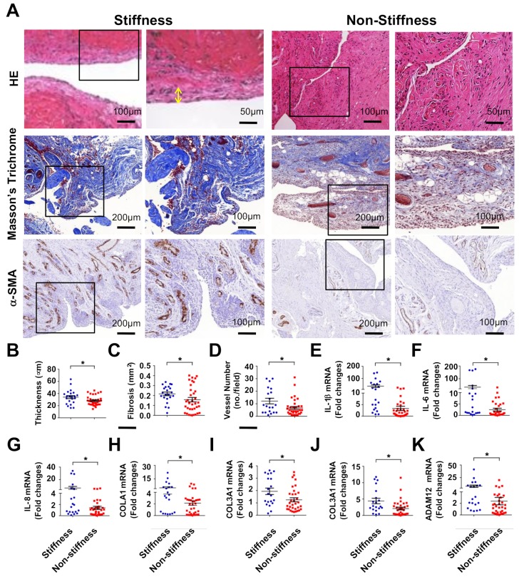
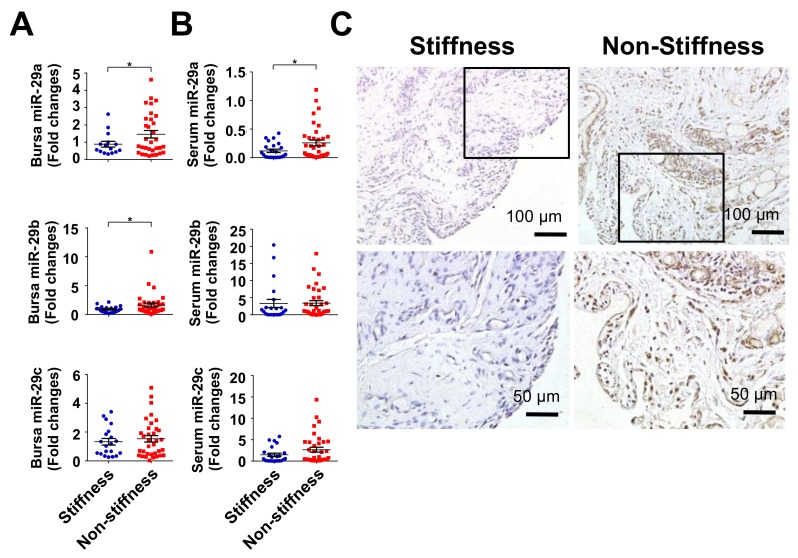
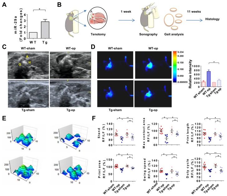
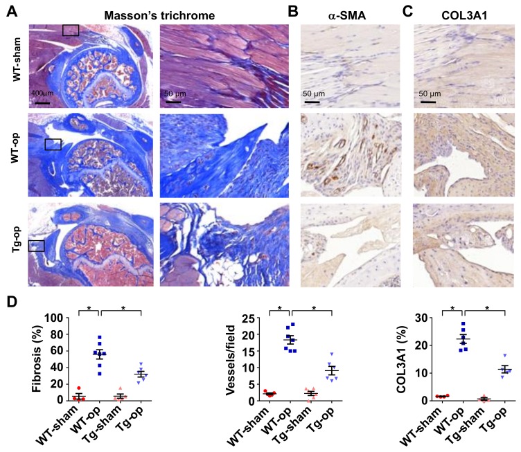
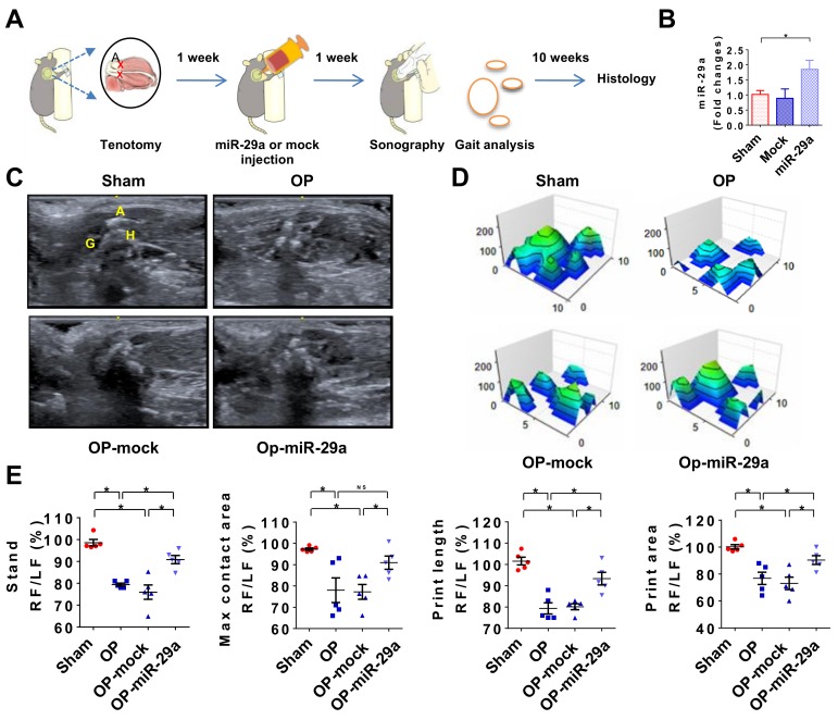
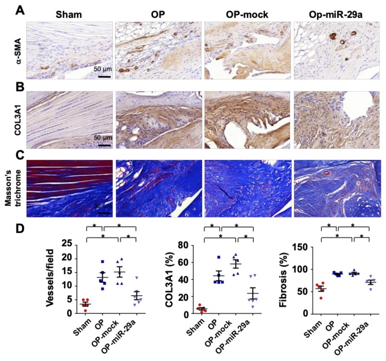
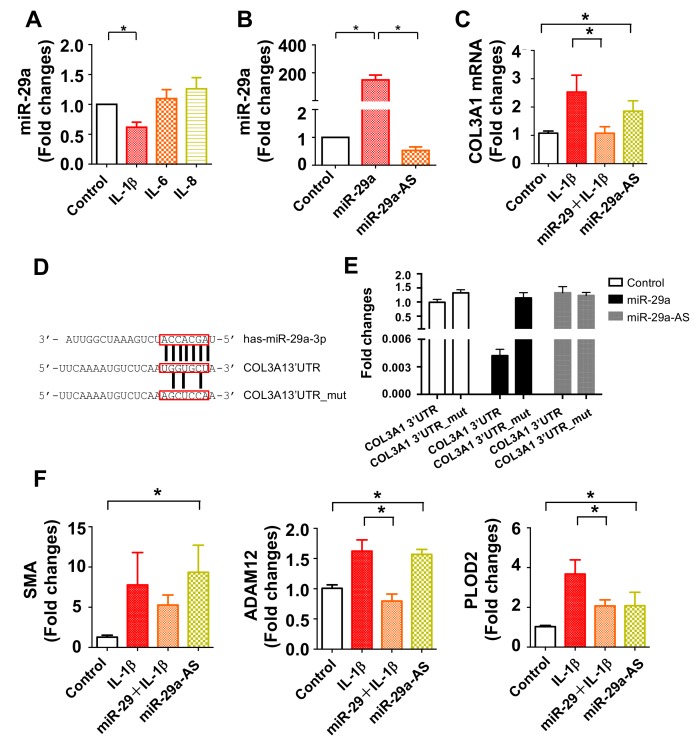
Rotator cuff lesion with shoulder stiffness is a major cause of shoulder pain and motionlessness. Subacromial bursa fibrosis is a prominent pathological feature of the shoulder disorder. MicroRNA-29a (miR-29a) regulates fibrosis in various tissues; however, the miR-29a action to subacromial bursa fibrosis remains elusive. Here, we reveal that subacromial synovium in patients with rotator cuff tear with shoulder stiffness showed severe fibrosis, hypertrophy, and hyperangiogenesis histopathology along with significant increases in fibrotic matrices collagen (COL) 1A1, 3A1, and 4A1 and inflammatory cytokines, whereas miR-29a expression was downregulated. Supraspinatus and infraspinatus tenotomy-injured shoulders in transgenic mice overexpressing miR-29a showed mild swelling, vascularization, fibrosis, and regular gait profiles as compared to severe rotator cuff damage in wild-type mice. Treatment with miR-29a precursor compromised COL3A1 production and hypervascularization in injured shoulders. In vitro, gain of miR-29a function attenuated COL3A1 expression through binding to the 3'-untranslated region (3'-UTR) of COL3A1 in inflamed tenocytes, whereas silencing miR-29a increased the matrix expression. Taken together, miR-29a loss is correlated with subacromial bursa inflammation and fibrosis in rotator cuff tear with shoulder stiffness. miR-29a repressed subacromial bursa fibrosis through directly targeting COL3A1 mRNA, improving rotator cuff integrity and shoulder function. Collective analysis offers a new insight into the molecular mechanism underlying rotator cuff tear with shoulder stiffness. This study also highlights the remedial potential of miR-29a precursor for alleviating the shoulder disorder.

Keywords: fibrosis; miR-29a; shoulder stiffness; subacromial bursa.


Conflict of interest statement

The authors declare no conflict of interest.

Figures

Figure 1
Figure 1
Analysis of fibrosis histopathology and fibrotic matrix expression of subacromial bursa in patients with rotator cuff lesion with or without stiffness. Subacromial bursa in the stiffness group show hypertrophy (yellow two-way arrow) as evident from hematoxylin and eosin (HE) staining. Masson’s trichrome staining displayed spacious fibrotic tissue (blue) rather than muscle tissue (red) in the stiffness group. Strong α-SMA immunostaining (brown) is exhibited in the stiffness group (A). The boxes stand for selected regions of interest for high power field images shown in panels 2 and 4 Subacromial bursa membrane thickness (B), fibrotic tissue area (C), and vessel formation (D) were significantly increased in the stiffness group. Inflammatory cytokines IL-1β (E), IL-6 (F), IL-8 (G) along with fibrotic matrices COL1A1 (H), COL13A1 (I), COL4A1 (J), and ADAM12 (K) expression were significantly upregulated in the stiffness group. Data are expressed as mean ± standard error of mean (SEM) from 22 patients in the stiffness group and 35 patients in the non-stiffness group. Asterisks (*) indicate significant differences between stiffness and non-stiffness group analyzed using Wilcoxon test (p < 0.05). HE, hematoxylin and eosin; α-SMA, α-smooth muscle actin; IL, interleukin; COL, collagen.
Figure 2
Figure 2
Analysis of miR-29 expression in subacromial bursa and serum. miR-29a and miR-29b expression were decreased in subacromial bursa in the stiffness group (A). Serum miR-29a rather than miR-29b and miR-29c levels were significantly downregulated in the stiffness group (B). Cells within subacromial bursa specimens in the stiffness group displayed weak miR-29a transcripts (brown) as compared to the non-stiffness group as evident from in situ hybridization (C). The boxes stand for selected regions of interest for high-power field images shown in right panels. Data are expressed as mean ± SEM from 22 patients with stiffness and 35 patients without stiffness. Asterisks (*) indicate significant differences between stiffness and non-stiffness group analyzed using Wilcoxon test (p < 0.05).
Figure 3
Figure 3
Analysis of rotator cuff lesion and gait profiles of wild-type mice and miR-29aTg mice. miR-29a expression was significantly increased in miR-29aTg mice (A). Schematic drawings for surgery-induced rotator cuff injury in mice (B). Echogenicity of injured shoulders was compromised in miR-29aTg mice (C). Inflammation in the injured tendon as evident from fluorescence 2-deoxyglucose uptake was downregulated in miR-29aTg mice (D). The tenotomy-mediated irregular footprint histograms (E) along with decreased stand time, maximum contact area, footprint length, footprint area, swing speed, and duty cycle (F) of forelegs with injured shoulders were improved in miR-29aTg mice. Data are expressed as mean ± SEM from 6–9 mice analyzed using ANOVA test and Bonferroni post-hoc test. Asterisks * (p < 0.05) indicate significant difference between groups. A, acromion; G, glenoid; H, humeral head; Op, tenotomy surgery; RF, right forelimb; LF, left forelimb; NS, not significant.
Figure 4
Figure 4
Histological analysis of rotator cuff injury in wild-type mice and miR-29aTg mice. Injured tendon in miR-29aTg mice showed moderate response to the tenotomy-mediated fibrotic tissue formation (blue) as evident from Masson’s trichrome staining (A). Boxes stand for selected regions of interest for high-power field images shown in right panels. The injured sites in miR-29aTg mice showed weak α-SMA immunostaining (B) and COL3A1 immunostaining (brown) (C). miR-29a overexpression significantly improved fibrotic tissue area, vessel number and COL3A1 production (D). Data are expressed as mean ± SEM from 5–7 mice analyzed using ANOVA test and Bonferroni post-hoc test. Asterisks * indicate significant difference between groups (p < 0.05).
Figure 5
Figure 5
miR-29a precursor treatment improvement of shoulder injury. Schematic drawings for miR-29a precursor treatment for shoulder injury (A). miR-29a expression was increased at 1 week upon miR-29a precursor treatment (B). Echogenicity of injured shoulder was compromised upon miR-29a precursor treatment (C). Tenotomy-mediated irregular footprint (D) along with stand time, maximum contact area, footprint length, and footprint area (E) were improved upon miR-29a precursor treatment. Data are expressed as mean ± SEM from 5–6 mice. Asterisks * (p < 0.05) indicate significant difference between groups analyzed using ANOVA test and Bonferroni post-hoc test. A, acromion; G, glenoid; H, humeral head; Op, tenotomy surgery; RF, right forelimb; LF, left forelimb; NS, not significant.
Figure 6
Figure 6
Histopathological analysis of injured shoulders with or without miR-29a precursor treatment. Injured sites in the miR-29a-treated group showed moderate α-SMA immunostaining (brown) (A) and COL3A1 immunoreaction (brown) (B) along with mild fibrosis tissue formation (blue) as evident from Masson’s trichrome staining (C). The tenotomy upregulation of vessel formation, COL3A1 production and fibrotic tissue area were compromised upon miR-29a precursor treatment (D). Data are expressed as mean ± SEM from 5–6 mice analyzed using ANOVA test and Bonferroni post-hoc test. Asterisks * indicate significant difference between groups (p < 0.05).
Figure 7
Figure 7
Analysis of miR-29a action to human tenocytes. IL-1β rather than IL-6 and IL-8 significantly decreased miR-29a expression in tenocytes. Cell cultures were incubated in 1 ng/mL IL-1β, IL-6, or IL-8 for 24 h (A). miR-29a expression was increased in tenocytes upon miR-29a precursor transfection, whereas miR-29a antisense oligonucleotide (miR-29a-AS) transfection reduced the expression (B). miR-29 precursor transfection attenuated the IL-1β-mediated COL3A1 expression. miR-29a knockdown alone provoked COL3A1 expression (C). Sequence (red boxes) of the miR-29a targeted 3′-untranalated region (3′-UTR) and 3-base pair mutant of COL3A1 were constructed for luciferase reporter assay (D). miR-29a precursor transfection decreased luciferase reporter activity of 3′-UTR rather than mutant of COL3A1 in tenocytes, whereas miR-29a-AS transfer increased the luciferase reporter activity (E). miR-29a precursor attenuated the IL-1b-augmented SMA, ADAM12, and PLOD2 expression. miR-29a-AS increased fibrosis marker expression in tenocytes (F). Data are expressed as mean ± SEM calculated from three experiments analyzed using ANOVA test and Bonferroni post-hoc test. Asterisks * indicate significant difference between groups (p < 0.05).

Similar articles

Cited by

References

    1. Thomopoulos S., Parks W.C., Rifkin D.B., Derwin K.A. Mechanisms of tendon injury and repair. J. Orthop. Res. 2015;33:832–839. doi: 10.1002/jor.22806. - DOI - PMC - PubMed
    1. Zhang K., de Sa D., Kanakamedala A., Sheean A.J., Vyas D. Management of Concomitant Preoperative Rotator Cuff Pathology and Adhesive Capsulitis: A Systematic Review of Indications, Treatment Approaches, and Outcomes. Arthroscopy. 2019;35:979–993. doi: 10.1016/j.arthro.2018.10.126. - DOI - PubMed
    1. Blaine T.A., Cote M.A., Proto A., Mulcahey M., Lee F.Y., Bigliani L.U. Interleukin-1beta stimulates stromal-derived factor-1alpha expression in human subacromial bursa. J. Orthop. Res. 2011;29:1695–1699. doi: 10.1002/jor.21416. - DOI - PubMed
    1. Harrison A.K., Flatow E.L. Subacromial impingement syndrome. J. Am. Acad. Orthop. Surg. 2011;19:701–708. doi: 10.5435/00124635-201111000-00006. - DOI - PubMed
    1. Carr A.J., Murphy R., Dakin S.G., Rombach I., Wheway K., Watkins B., Franklin S.L. Platelet-Rich Plasma Injection with Arthroscopic Acromioplasty for Chronic Rotator Cuff Tendinopathy: A Randomized Controlled Trial. Am. J. Sports Med. 2015;43:2891–2897. doi: 10.1177/0363546515608485. - DOI - PubMed