Review
. 2019 Dec;40(12):1163-1179.
doi: 10.1016/j.it.2019.10.002. Epub 2019 Nov 12.

Foam Cells: One Size Doesn't Fit All

Affiliations
Review

Foam Cells: One Size Doesn't Fit All

Valentina Guerrini et al. Trends Immunol. 2019 Dec.

Abstract

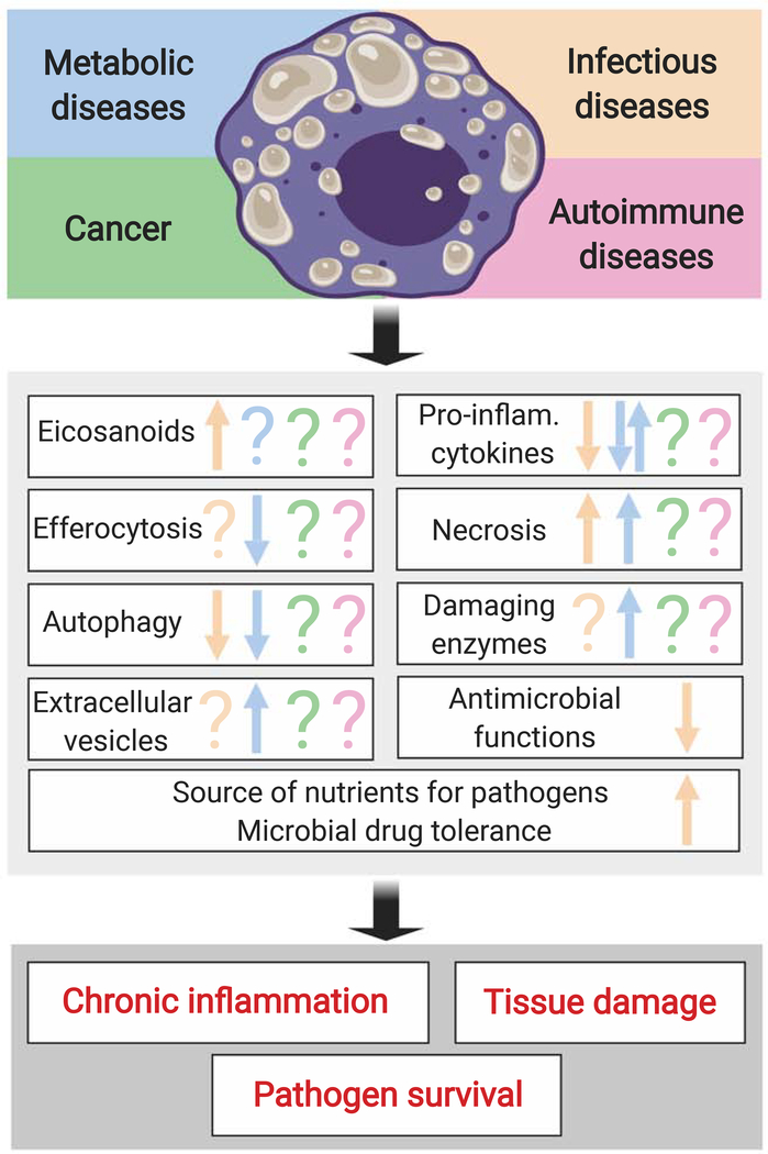
Chronic inflammation in many infectious and metabolic diseases, and some cancers, is accompanied by the presence of foam cells. These cells form when the intracellular lipid content of macrophages exceeds their capacity to maintain lipid homeostasis. Concurrently, critical macrophage immune functions are diminished. Current paradigms of foam cell formation derive from studies of atherosclerosis. However, recent studies indicate that the mechanisms of foam cell biogenesis during tuberculosis differ from those operating during atherogenesis. Here, we review how foam cell formation and function vary with disease context. Since foam cells are therapeutic targets in atherosclerosis, further research on the disease-specific mechanisms of foam cell biogenesis and function is needed to explore the therapeutic consequences of targeting these cells in other diseases.

Keywords: atherosclerosis; chronic inflammation; foam cells; lipid droplets; macrophage; tuberculosis.


Figures

Figure 1.
Figure 1.. Foam cells can contribute to disease pathogenesis.
The top panel shows certain types of human diseases associated with the presence of foam cells. The middle panel lists the macrophage functions that have been studied in foam cells. The bottom panel indicates the major, disease-promoting outcomes associated with the maladaptive, foam-cell responses. Arrow up, upregulation; arrow down, downregulation; question mark, unknown. Inflam: inflammatory.
Key Figure, Figure 2.
Key Figure, Figure 2.. Human tuberculous granulomas and atheromas show similar architecture but different foam cell biogenesis.
(A, B) Lesional structure and cellular composition of human necrotizing tuberculous granulomas (left panels) and advanced atherosclerotic lesions (right panels) (haematoxylin and eosin (H&E) staining). (A) Left: Necrotic core (caseum, C) and cellular (CR) regions of granuloma. Higher magnification (black box inset) shows the foam-cell-rich area (arrows). Right: Atherosclerotic plaque in coronary artery. The bluish discoloration (*) within the necrotic core (NC) is due to inflammatory cells. At higher magnification, the foam-cell-rich area (arrows) is located near necrotic areas; the inflamed plaque shows surface erosion and luminal thrombus (Th). In both panels, the presence of foam cells shows as vacuole-rich areas due to lipid loss during H&E staining. Scale bars are shown as available. Images were reproduced with permission [2, 119]. (B) The two lesions have similar architectures and share immune and stromal cell types, albeit triggered by different stimuli, located in different anatomical compartments, and having different geometry (quasi-symmetric lesion with central necrotic core in tuberculosis; asymmetric lesion with lateral necrotic core in atherosclerosis). Mtb, M. tuberculosis. (C) Tuberculous and atherogenic foam cell biogenesis. Left: Bacteria-activated TLR2 signaling induces the transcription factor PPARγ. Macrophages secrete TNFα and 3-hydroxy butyrate (3HB). TNF-receptor signaling triggers caspase and mTORC1 signaling [2], inducing triglyceride (TAG) synthesis and blocking TAG degradation [76]. 3HB binds the G-protein-coupled receptor GPR109A and prevents TAG hydrolysis by stabilizing perilipins [70]. TAG accumulation is also induced by micro-RNA33 (miRNA33) (hydrolysis inhibition) and the transcription factor TR4 (unknown mechanism). Right: Atherogenic foam cells are generated by inducing cholesterol-rich lipoprotein uptake, subverting cholesterol trafficking, and reducing cholesterol efflux [62]. The uncontrolled uptake of native LDL and modified LDL (mLDL) (mediated by scavenger receptors CD36, SR-A, LOX-1) disrupts cholesterol homeostasis [83, 84, 120]. Excess free cholesterol (FC) is accumulated as cholesteryl esters (CE) in lipid droplets [85]. CE mobilization through lipophagy and lypolysis is impaired and FC efflux through ABCA1 and ABCG1 transporters is decreased [51, 89, 92]. Arrowheads vs Barheads = positive vs negative regulation.

References

    1. Childs BG et al. (2016) Senescent intimal foam cells are deleterious at all stages of atherosclerosis. Science 354 (6311), 472–477. - PMC - PubMed
    1. Guerrini V et al. (2018) Storage lipid studies in tuberculosis reveal that foam cell biogenesis is disease-specific. PLoS Pathog 14 (8), e1007223. - PMC - PubMed
    1. Grajchen E et al. (2018) The physiology of foamy phagocytes in multiple sclerosis. Acta Neuropathol Commun 6 (1), 124. - PMC - PubMed
    1. Ye H et al. (2018) Tumor-associated macrophages promote progression and the Warburg effect via CCL18/NF-kB/VCAM-1 pathway in pancreatic ductal adenocarcinoma. Cell Death Dis 9 (5), 453. - PMC - PubMed
    1. Christiani DC (2019) Vaping-Induced Lung Injury. N Engl J Med. - PubMed

Publication types