. 2019 Dec 15:239:117053.
doi: 10.1016/j.lfs.2019.117053. Epub 2019 Nov 13.

Intracardiac administration of ephrinA1-Fc preserves mitochondrial bioenergetics during acute ischemia/reperfusion injury

Affiliations

Intracardiac administration of ephrinA1-Fc preserves mitochondrial bioenergetics during acute ischemia/reperfusion injury

Maria J Torres et al. Life Sci. .

Abstract

Aims: Intracardiac injection of recombinant EphrinA1-Fc immediately following coronary artery ligation in mice reduces infarct size in both reperfused and non-reperfused myocardium, but the cellular alterations behind this phenomenon remain unknown.

Main methods: Herein, 10 wk-old B6129SF2/J male mice were exposed to acute ischemia/reperfusion (30minI/24hrsR) injury immediately followed by intracardiac injection of either EphrinA1-Fc or IgG-Fc. After 24 h of reperfusion, sections of the infarct margin in the left ventricle were imaged via transmission electron microscopy, and mitochondrial function was assessed in both permeabilized fibers and isolated mitochondria, to examine mitochondrial structure, function, and energetics in the early stages of repair.

Key findings: At a structural level, EphrinA1-Fc administration prevented the I/R-induced loss of sarcomere alignment and mitochondrial organization along the Z disks, as well as disorganization of the cristae and loss of inter-mitochondrial junctions. With respect to bioenergetics, loss of respiratory function induced by I/R was prevented by EphrinA1-Fc. Preservation of cardiac bioenergetics was not due to changes in mitochondrial JH2O2 emitting potential, membrane potential, ADP affinity, efficiency of ATP production, or activity of the main dehydrogenase enzymes, suggesting that EphrinA1-Fc indirectly maintains respiratory function via preservation of the mitochondrial network. Moreover, these protective effects were lost in isolated mitochondria, further emphasizing the importance of the intact cardiomyocyte ultrastructure in mitochondrial energetics.

Significance: Collectively, these data suggest that intracardiac injection of EphrinA1-Fc protects cardiac function by preserving cardiomyocyte structure and mitochondrial bioenergetics, thus emerging as a potential therapeutic strategy in I/R injury.

Keywords: Cardioprotection; Mitochondrial bioenergetics; Myocardial infarction; ephrinA1.


Conflict of interest statement

Conflict of Interest

A conflicting interest exists when professional judgment concerning a primary interest (such as patient’s welfare or the validity of research) may be influenced by a secondary interest (such as financial gain or personal rivalry). It may arise for the authors when they have financial interest that may influence their interpretation of their results or those of others. Examples of potential conflicts of interest include employment, consultancies, stock ownership, honoraria, paid expert testimony, patent applications/registrations, and grants or other funding.

Figures

Figure 1.
Figure 1.. Experimental Model.
Graphical representation of left anterior descending coronary artery (LAD) ligation depicting EA1/IgG intracardiac injection at border zone diffusing toward apex and transmurally as well as the allocation of heart sections to the respective experimental endpoints. Two slices in the transversal plane were collected for 1. transmission electron microscopy (TEM) and preparation of permeabilized fibers and isolated mitochondria, and 2. metabolite analysis using UPLC and enzyme activity analyses.
Figure 2.
Figure 2.. Protein levels of EphrinA1 (A), SERCA2a (B), chchd3 (C) and GRP78 (D) by western blotting.
Representative blots are shown in the bottom. Bars are means +/− SEM and normalized to GAPDH levels. * p<0.05, ** p<0.005, and *** p<0.0005 vs sham, and # p<0.05, ### p<0.0005 vs I/R from one-way ANOVA analysis. n=4 mice/group.
Figure 3.
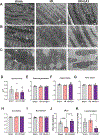
Figure 3.. EA1 administration in I/R helps preserve cardiomyocyte and mitochondrial 2-D ultrastructure.
(A–C) Representative transmission electron microscopy (TEM) images from a section of left ventricle border infarct in sham, and I/R+/−EA1, at x12,000 (A), x20,000 (B), and x40,000 (c) magnitude. Images captured with an EMSIS MegaView G3 charge-coupled device digital camera. Scale bars: 2 μm in (A), 1 μm in (B) and 0.5 μm in (D). (D–I) Analysis of morphological parameters in mitochondria (expressed in appropriate dimensions or arbitrary units): surface area (D), external perimeter (E), aspect ratio (F), form factor (G), circularity (H), roundness (I), computed as described in methods. (J) Number of electron-dense inter-mitochondrial junctions, and (K) lipid droplets. Bars are means +/− SEM from averages of up to 18 images/mouse, n=3 for sham, and n=4–5 for I/R +/− EA1. *p<0.05 from one-way ANOVA analysis.
Figure 4.
Figure 4.. Evaluation of mitochondrial dynamics.
Western blot analysis of Beclin1 (A), Drp1 (B), Fis1 (C), Opa1 (D), and Mfn1 (E). Representative blots are shown in the bottom. Bars are means +/− SEM and normalized to GAPDH levels. * p<0.05 vs sham, and # p<0.05 vs I/R from one-way ANOVA analysis. n=4 mice/group.
Figure 5.
Figure 5.. EA1 preserves mitochondrial bioenergetics in cardiac PmFBs.
(A) Mitochondrial respiratory capacity measured in permeabilized fibers from left ventricle. Substrates added sequentially: 18 μM Palpmitoyl-carnitine (PC), 5 mM L-carnitine, 0.5 mM malate, 4 mM ADP, 10 mM pyruvate, 10 mM glutamate, 10 mM succinate. (B) Mitochondrial JH2O2 emitting potential measured after the addition of 10 mM succinate, and 1 μM BCNU + 1 μm auranufin (left panel), or 18 μM palmitoyl-carnitine, 5 mM L-carnitine and 1 μM BCNU + 1 μm auranufin (right panel). (C) Citrate synthase activity measured in homogenates of the PmFBs utilized in 3A and 3D. (D) Representative creatine kinase energetic clamp in PmFBs from LV after I/R +/− EA1. Steady-state JO2 was measured and ΔGATP was calculated as described elsewhere, after each addition of PCr [23]. (E) Force-flow relationship where the slope represents conductance and the x-intercept the static head (ΔGATP at JO2 = 0, represented at (F)). (G) ADP kinetics, showing data fitted to a Michaelis Menten function. (H) Eadie-Hofstee plot of the data presented in G, where y = −(KMADP)x + Vmax. Slopes (KM) were not statistically different. Bars are means ± SEM. * p<0.05, ** p<0.005 vs. sham and # p<0.05 vs. I/R from one-way ANOVA analysis. N = 6–8 mice/group.
Figure 6.
Figure 6.. EA1-mediated protective effects in mitochondrial bioenergetics are not detected in the isolated organelle.
(A) Enzymatic activities of key dehydrogenase enzymes in isolated mitochondria. Data are expressed as % of maximal rate value in each respective assay, n=3 mice/group. (B and C) Steady-state OXPHOS flux rates of O2 consumption (B) and ATP production (C) were determined simultaneously in real-time using a glucose / hexokinase / glucose-phosphate dehydrogenase respiratory clamp after the sequential addition of 20 and 200 μM ADP. Respiration was supported by 0.5 mM malate, 5 mM pyruvate, 5 mM glutamate, 5 mM succinate. (D) Resulting ATP/O ratio calculated from steady-state JATP/JO2. (E) Mitochondrial complex V enzymatic activity determined in isolated mitochondria. N=3 mice/group (F) Mitochondrial membrane potential determined using a TPP probe at different respiratory states via titration of malonate. (G) Mitochondrial respiratory capacity measured in isolated mitochondria from LV following the same protocol as in Figure 2A. Substrates added sequentially: 18 μM palmitoyl-carnitine (PC), 5 mM L-carnitine (carn), 0.5 mM malate (Mal), 4 mM ADP, 10 mM pyruvate (Pyr), 10 mM glutamate (glut), 10 mM succinate (succ). Values are means ± SEM, NS = no statistical differences found. N = 4 mice/group, with each data point representing a pooled sample from 2 mice.
Figure 7.
Figure 7.. Levels of metabolites determined by UPLC.
Guanosine triphosphate GTP (A), adenosine tri- (B), di-(C) and mono-phosphate (D), respective ATP/ADP (E) and ATP/AMP (F) ratios, adenine (G), hypoxanthine (H), IMP (I), and total nucleotides (J). Bars are means +/− SEM. No statistical differences were detected from a one-way ANOVA analysis. n = 9–15 mice/group.
Figure 8.
Figure 8.. EA1 administration in I/R preserves phosphorylation of α-Tubulin.
Western blot analysis of α-Tubulin (A) and (Tyr272) phosphorylated-α-Tubulin (B) in LV homogenates. (C) Calculated p-α-Tubulin/ α-Tubulin ratio from a and b. Representative blots are shown on the right. Bars are means ± SEM, * p<0.05, ** p<0.005 vs. sham and ## p<0.005, ### p<0.0005 vs. I/R from one-way ANOVA analysis. N = 4 mice/group.

References

    1. Benjamin EJ, Virani SS, Callaway CW, Chang AR, Cheng S, Chiuve SE et al. Heart Disease and Stroke Statistics-2018 Update: A Report From the American Heart Association. Circulation. 2018. doi:10.1161/CIR.0000000000000558. - DOI - PubMed
    1. Yellon DM, Hausenloy DJ. Myocardial reperfusion injury. N Engl J Med. 2007;357(11):1121–35. doi:10.1056/NEJMra071667. - DOI - PubMed
    1. Lassen JF, Botker HE, Terkelsen CJ. Timely and optimal treatment of patients with STEMI. Nat Rev Cardiol. 2013;10(1):41–8. doi:10.1038/nrcardio.2012.156. - DOI - PubMed
    1. Brown DA, Perry JB, Allen ME, Sabbah HN, Stauffer BL, Shaikh SR et al. Expert consensus document: Mitochondrial function as a therapeutic target in heart failure. Nat Rev Cardiol. 2017;14(4):238–50. doi:10.1038/nrcardio.2016.203. - DOI - PMC - PubMed
    1. Murphy E, Ardehali H, Balaban RS, DiLisa F, Dorn GW 2nd, Kitsis RN et al. Mitochondrial Function, Biology, and Role in Disease: A Scientific Statement From the American Heart Association. Circ Res. 2016;118(12):1960–91. doi:10.1161/RES.0000000000000104. - DOI - PMC - PubMed

MeSH terms