Review
. 2020 Apr:100:199-211.
doi: 10.1016/j.semcdb.2019.11.002. Epub 2019 Nov 13.

Convergent extension in mammalian morphogenesis

Affiliations
Review

Convergent extension in mammalian morphogenesis

Ann Sutherland et al. Semin Cell Dev Biol. 2020 Apr.

Abstract

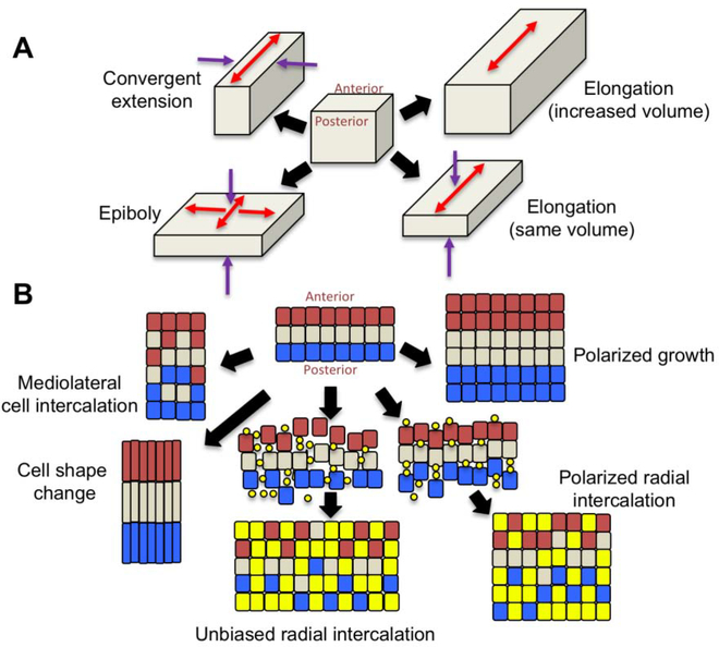
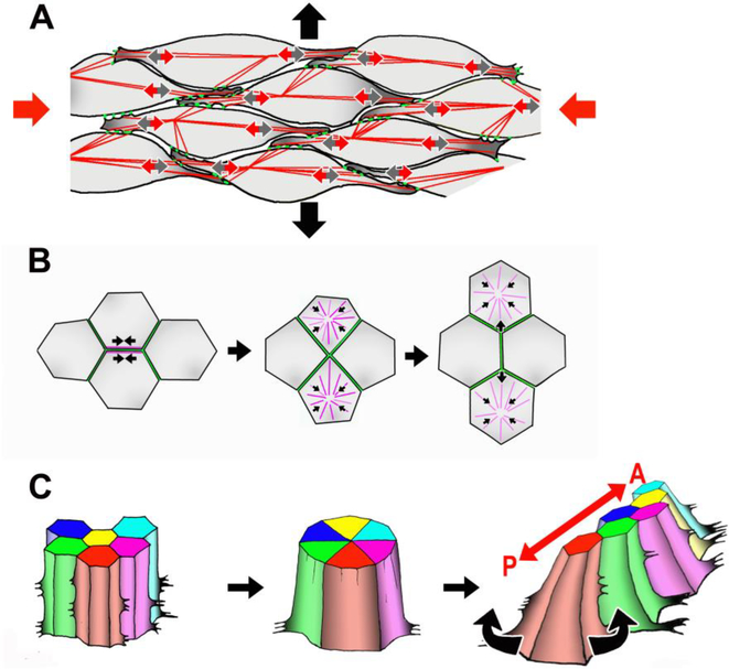
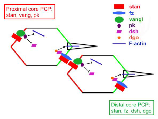
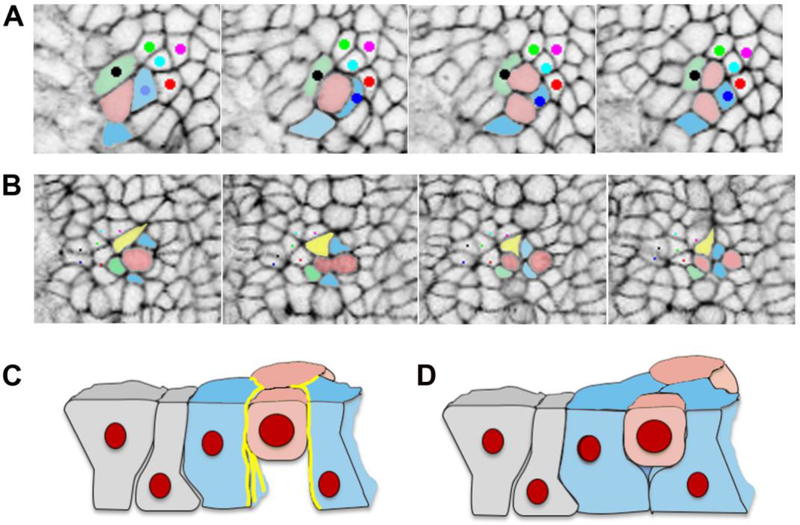
Convergent extension is a fundamental morphogenetic process that underlies not only the generation of the elongated vertebrate body plan from the initially radially symmetrical embryo, but also the specific shape changes characteristic of many individual tissues. These tissue shape changes are the result of specific cell behaviors, coordinated in time and space, and affected by the physical properties of the tissue. While mediolateral cell intercalation is the classic cellular mechanism for producing tissue convergence and extension, other cell behaviors can also provide similar tissue-scale distortions or can modulate the effects of mediolateral cell intercalation to sculpt a specific shape. Regulation of regional tissue morphogenesis through planar polarization of the variety of underlying cell behaviors is well-recognized, but as yet is not well understood at the molecular level. Here, we review recent advances in understanding the cellular basis for convergence and extension and its regulation.

Keywords: Axial elongation; Convergent extension; Gastrulation; Morphogenesis; Mouse; Planar cell polarity.


Figures

Figure 1.
Figure 1.. Tissue shape changes.
A. Ways in which a tissue can change shape. Convergent extension makes the tissue longer and narrower, epiboly makes it wider and flatter, and elongation makes the tissue longer without changing the width, and can either occur with a change in tissue volume or without. Arrows indicated the direction of tissue shape change (red = expansion; purple = narrowing or thinning). B. Cellular behaviors that can lead to the tissue shape changes shown in A. Convergent extension can be accomplished by polarized cell intercalation or by polarized cell shape change. Radial intercalation occurs when cells from an underlying layer insert themselves into the upper layer. In this example, yellow dots are cells starting to insert from underneath, and become the yellow squares as they join the overlying layer. Radial intercalation can be either unbiased, expanding the original cell layer in both anterior-posterior and mediolateral dimensions (epiboly), or polarized, expanding the tissue in only one dimension (elongation), and in both cases the tissue thins. Polarized cell division when coupled with cell growth leads to elongation with an increase in tissue volume.
Figure 2.
Figure 2.. Cellular behaviors underlying CE.
A. Mesenchymal cells extend mediolateral lamellipodial protrusions (small grey arrows), form attachments to neighboring cells (green dots), and contraction of the actomyosin cytoskeleton (red lines) exerts traction (small red arrows) to move the cell between its neighbors. In the illustrated case, this process provides a convergence force along the mediolateral axis (large red arrows) to drive extension along the A/P axis (large black arrows). B. T1 process of epithelial cell rearrangement. Actomyosin contraction along a boundary between two cells brings four cells together at a common vertex, and apical myosin contraction elongates a new boundary between the cells not formerly in contact. C. Rosette formation involves contraction of multiple boundaries to form a common vertex between 5–10 cells, followed by the formation of new boundaries along the opposite axis to create a longer, narrower array of cells. As pictured here, the basal ends of mouse neural cells are motile, and biased protrusive activity likely drives the polarity of the rearrangement [111].
Figure 3.
Figure 3.. stan/fz planar cell polarity signaling pathway in Drosophila.
Polarized distribution of core planar polarity proteins within epithelial cells.
Figure 4.
Figure 4.. Cell division generating cell rearrangement in the mouse neural plate.
A. In this example, the pink cell divides, and the daughter cells stay in contact, while one pink daughter cell separates the blue and green cells. B. In this example the pink cell divides, and the two blue cells insert themselves between the two pink daughter cells, and one pink daughter cell separates the yellow and green cells. C and D. Schematics of the behavior of the basal ends of neighboring cells during cell division, based on published data for chick epiblast [115]. C. When actin and myosin localization (yellow lines) is strong and cadherin low at the lateral boundaries of cells neighboring a mitotic cell, these cells do not fill in the space basal to the mitotic cell to form new intercellular junctions, and as a result do not separate the daughter cells. D. When actin and myosin are not strongly localized at the lateral boundaries of cells neighboring a mitotic cell, these cells are able to fill in the space basal to the mitotic cell, forming new junctions and separating the daughter cells.
Figure 5.
Figure 5.. Initiation of coiling in the Wolffian duct.
Wolffian ducts collected from hoxb7 GFP transgenic mice at embryonic day (E)14.5, E16.5, E18.5, and postnatal (P) day 1 (P1). GFP is expressed in the epithelial cells of the ducts, allowing the process of duct coiling to be appreciated. Coiling proceeds from proximal to distal, and is initially two-dimensional at E16.5, and three-dimensional by E18.5. Republished with permission of John Wiley and Sons, Inc., from Hinton et al [174]; permission conveyed through Copyright Clearance Center, Inc.

References

    1. Nishimura T, Honda H, Takeichi M, Planar cell polarity links axes of spatial dynamics in neural-tube closure, Cell. 149 (2012) 1084–1097. doi:10.1016/j.cell.2012.04.021. - DOI - PubMed
    1. Condic ML, Fristrom D, Fristrom JW, Apical cell shape changes during Drosophila imaginal leg disc elongation: A novel morphogenetic mechanism, Development. 111 (1991) 23–33. - PubMed
    1. Priess JR, Hirsh DI, Caenorhabditis elegans morphogenesis: The role of the cytoskeleton in elongation of the embryo, Dev. Biol 117 (1986) 156–173. doi:10.1016/0012-1606(86)90358-1. - DOI - PubMed
    1. Simske JS, Hardin J, Getting into shape: Epidermal morphogenesis in Caenorhabditis elegans embryos, BioEssays. 23 (2001) 12–23. doi:10.1002/1521-1878(200101)23:1<12::AID-BIES1003>3.0.CO;2-R. - DOI - PubMed
    1. Fischer E, Legue E, Doyen A, Nato F, Nicolas J-F, Torres V, Yaniv M, Pontoglio M, Defective planar cell polarity in polycystic kidney disease, Nat Genet. 38 (2006) 21–23. doi:10.1038/ng1701. - DOI - PubMed

Publication types

LinkOut - more resources