

Conjunctival goblet cells: Ocular surface functions, disorders that affect them, and the potential for their regeneration
- PMID: 31734511
- PMCID: PMC7004882
- DOI: 10.1016/j.jtos.2019.11.005
Conjunctival goblet cells: Ocular surface functions, disorders that affect them, and the potential for their regeneration
Erratum in
-
Corrigendum to "Conjunctival goblet cells: Ocular surface functions, disorders that affect them, and the potential for their regeneration" [Ocul Surf 18 (2019) 19-26].Ocul Surf. 2022 Jan;23:224. doi: 10.1016/j.jtos.2020.02.010. Epub 2020 Feb 29. Ocul Surf. 2022. PMID: 32120006 No abstract available.
Abstract
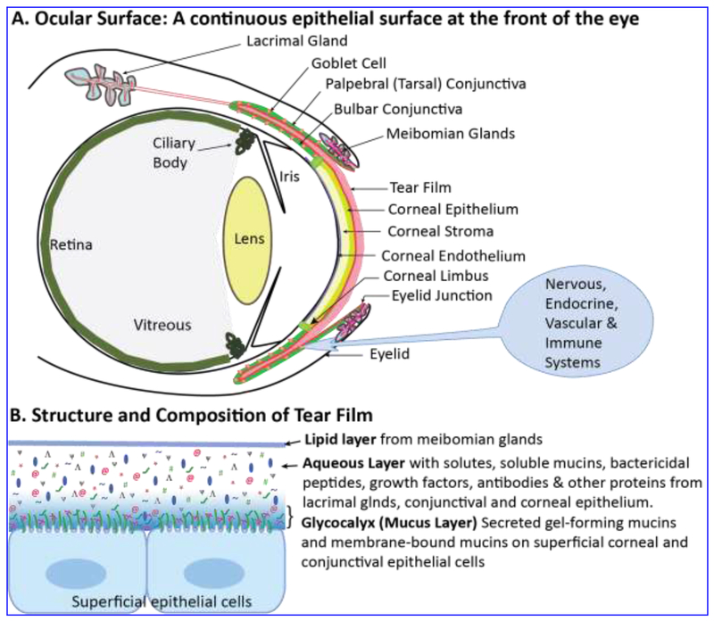
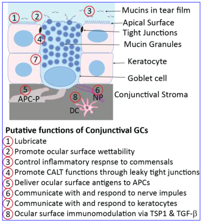
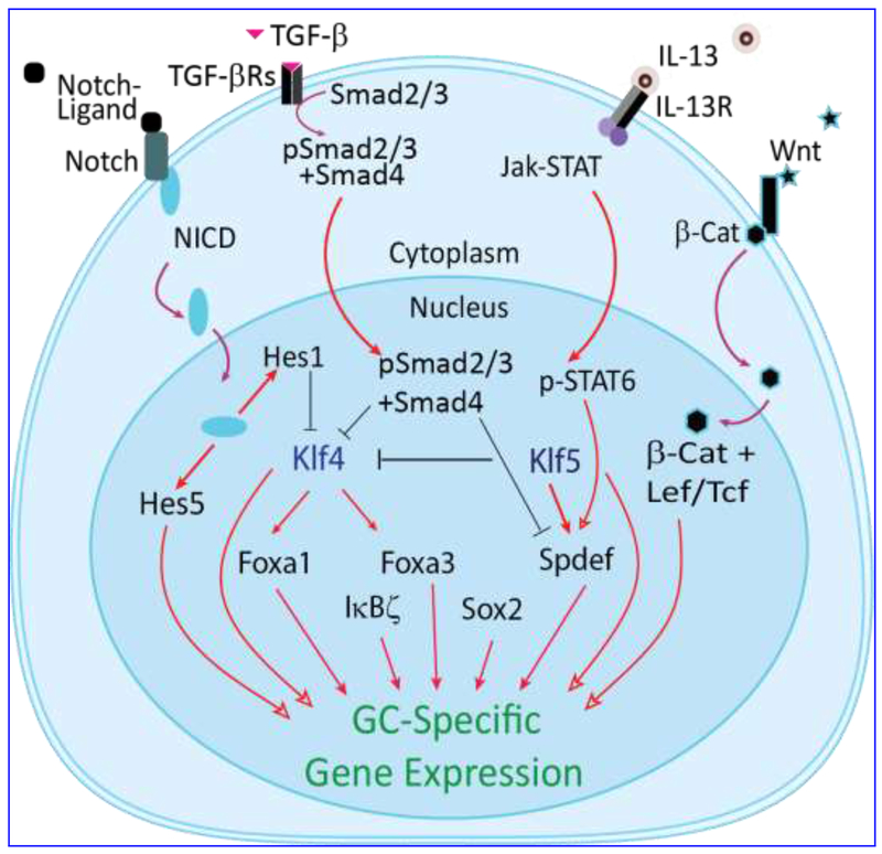
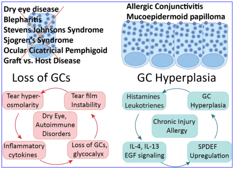
Conjunctival goblet cells (CGCs) are specialized cells that produce and secrete soluble mucins to the tear film that bathes the ocular surface. CGC numbers and functions are affected in various ocular surface diseases including dry eye disease with diverse etiologies. In this review we will (i) summarize the important functions of CGCs in ocular surface health, (ii) describe the ocular surface diseases that affect CGC numbers and function, (iii) provide an update on recent research outcomes that elucidate CGC differentiation, gene expression and functions, and (iv) present evidence in support of the prediction that restoring CGC numbers and/or functions is a viable strategy for alleviating ocular surface disorders that impact the CGCs.
Keywords: Conjunctiva; Cornea; Goblet cells; Ocular surface; Tear film.
Copyright © 2019 Elsevier Inc. All rights reserved.
Conflict of interest statement
Declaration of competing interest AW is a founder and holder of equity in Ocugenix, a privately held biotechnology start up that is developing activators of CXCR3 for the treatment of ocular pathologies. AW is a named inventor on patents held by the University of Pittsburgh on these potential therapeutics.
Figures
Similar articles
-
Resolvin D2 uses multiple Ca2+ -dependent signaling pathways to stimulate mucin secretion in rat and human conjunctival goblet cells.J Cell Physiol. 2022 Oct;237(10):3816-3833. doi: 10.1002/jcp.30854. Epub 2022 Sep 6. J Cell Physiol. 2022. PMID: 36066128 Free PMC article.
-
Imaging assessment of conjunctival goblet cells in dry eye disease.Clin Exp Ophthalmol. 2024 Jul;52(5):576-588. doi: 10.1111/ceo.14379. Epub 2024 Mar 30. Clin Exp Ophthalmol. 2024. PMID: 38553944 Review.
-
Reconsidering the central role of mucins in dry eye and ocular surface diseases.Prog Retin Eye Res. 2019 Jul;71:68-87. doi: 10.1016/j.preteyeres.2018.11.007. Epub 2018 Nov 22. Prog Retin Eye Res. 2019. PMID: 30471351 Review.
-
Surgery of the conjunctiva.Dev Ophthalmol. 2008;41:138-158. doi: 10.1159/000131086. Dev Ophthalmol. 2008. PMID: 18453766 Review.
-
Secreted Mucins on the Ocular Surface.Invest Ophthalmol Vis Sci. 2018 Nov 1;59(14):DES151-DES156. doi: 10.1167/iovs.17-23623. Invest Ophthalmol Vis Sci. 2018. PMID: 30481820 Review.
Cited by
-
Goblet Cell Differentiation Potential in Human Corneal Limbal Epithelial Progenitor Cells In Vitro.Invest Ophthalmol Vis Sci. 2020 Oct 1;61(12):27. doi: 10.1167/iovs.61.12.27. Invest Ophthalmol Vis Sci. 2020. PMID: 33112944 Free PMC article.
-
Suppression of NLRP3/Caspase-1/GSDMD Mediated Corneal Epithelium Pyroptosis Using Melatonin-Loaded Liposomes to Inhibit Benzalkonium Chloride-Induced Dry Eye Disease.Int J Nanomedicine. 2023 May 9;18:2447-2463. doi: 10.2147/IJN.S403337. eCollection 2023. Int J Nanomedicine. 2023. PMID: 37192892 Free PMC article.
-
Evaluation of ocular surface inflammation and systemic conditions in patients with systemic lupus erythematosus: a cross-sectional study.BMC Ophthalmol. 2024 Nov 12;24(1):492. doi: 10.1186/s12886-024-03760-8. BMC Ophthalmol. 2024. PMID: 39533209 Free PMC article.
-
The Role of Scanning Electron Microscopy in the Evaluation of Conjunctival Microvilli as an Early Biomarker of Ocular Surface Health: A Literature Review.J Clin Med. 2024 Dec 12;13(24):7569. doi: 10.3390/jcm13247569. J Clin Med. 2024. PMID: 39768491 Free PMC article. Review.
-
Thermoresponsive antioxidant metal-free carbon nanodot hydrogel: An effective therapeutic approach for ocular surface disease.Sci Adv. 2025 Jul 25;11(30):eadt8775. doi: 10.1126/sciadv.adt8775. Epub 2025 Jul 25. Sci Adv. 2025. PMID: 40712015 Free PMC article.
References
-
- Dartt DA. Regulation of mucin and fluid secretion by conjunctival epithelial cells. Prog Retin Eye Res 2002;21:555–576. - PubMed
-
- Farrand KF, Fridman M, Stillman IO, Schaumberg DA. Prevalence of Diagnosed Dry Eye Disease in the United States Among Adults Aged 18 Years and Older. Am J Ophthalmol 2017;182:90–98. - PubMed
-
- Stapleton F, Alves M, Bunya VY, et al. TFOS DEWS II Epidemiology Report. Ocul Surf 2017;15:334–365. - PubMed
Publication types
MeSH terms
Grants and funding
LinkOut - more resources
Full Text Sources
