

Resting state network plasticity related to picture naming in low-grade glioma patients before and after resection
- PMID: 31734532
- PMCID: PMC6861733
- DOI: 10.1016/j.nicl.2019.102010
Resting state network plasticity related to picture naming in low-grade glioma patients before and after resection
Abstract
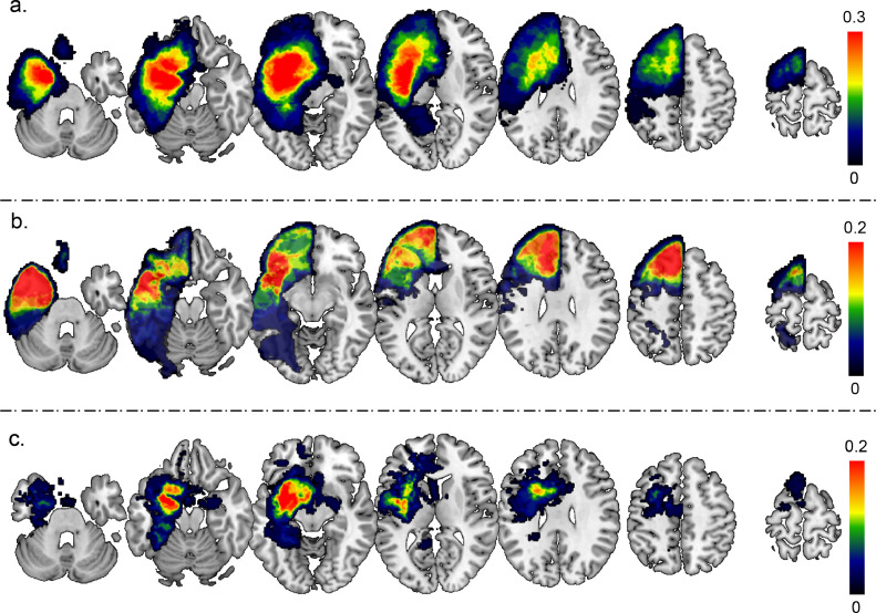
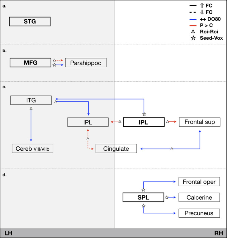
The dynamic connectome perspective states that brain functions arise from the functional integration of distributed and/or partly overlapping networks. Diffuse low-grade gliomas (DLGG) have a slow infiltrating character. Here we addressed whether and how anatomical disconnection following DLGG growth and resection might interfere with functional resting-state connectivity, specifically in relation to picture naming. Thirty-nine native French persons with a left DLGG were included. All underwent awake surgical resection of the tumor using direct brain electrostimulation to preserve critical eloquent regions. The anatomical disconnectivity risk following the DLGG volume and the resection, and the functional connectivity of resting-state fMRI images in relation to picture naming were evaluated prior to and three months after surgery. Resting-state connectivity patterns were compared with nineteen healthy controls. It was demonstrated that picture naming was strongly dependent on the semantic network that emerged from the integration and interaction of regions within multiple resting-state brain networks, in which their specific role could be explained in the light of the broader resting-state network they take part in. It emphasized the importance of a whole brain approach with specific clinical data input, during resting-state analysis in case of lesion. Adaptive plasticity was found in secondary regions, functionally connected to regions close to the tumor and/or cavity, marked by an increased connectivity of the right and left inferior parietal lobule with the left inferior temporal gyrus. In addition, an important role was identified for the superior parietal lobe, connected with the frontal operculum, suggesting functional compensation by means of attentional resources in order to name a picture via recruitment of the frontoparietal attention network.
Keywords: Connectivity; Glioma; Neurosurgery; Picture naming; Plasticity; Resting state.
Copyright © 2019. Published by Elsevier Inc.
Conflict of interest statement
None.
Figures
References
-
- Abel S, Dressel K, Kummerer D. Correct and errorneous picture naming responses in healthy subjects. Neurosci. Lett. 2009;463:167–171. - PubMed
-
- Aguirre GK, Detre JA, Alsop DC, D'Esposito M. The parahippocampus subserves topographical learning in man. Cereb. Cortex. 1996;6:823–829. - PubMed
-
- Almairac F, Herbet G, Moritz-Gasser S, de Champfleur NM, Duffau H. The left inferior fronto-occipital fasciculus subserves language semantics: a multilevel lesion study. Brain Struct. Funct. 2015;220:1983–1995. - PubMed
Publication types
MeSH terms
LinkOut - more resources
Full Text Sources
Medical
