. 2019 Dec:50:366-378.
doi: 10.1016/j.ebiom.2019.10.059. Epub 2019 Nov 14.

H19 promote calcium oxalate nephrocalcinosis-induced renal tubular epithelial cell injury via a ceRNA pathway

Affiliations

H19 promote calcium oxalate nephrocalcinosis-induced renal tubular epithelial cell injury via a ceRNA pathway

Haoran Liu et al. EBioMedicine. 2019 Dec.

Abstract

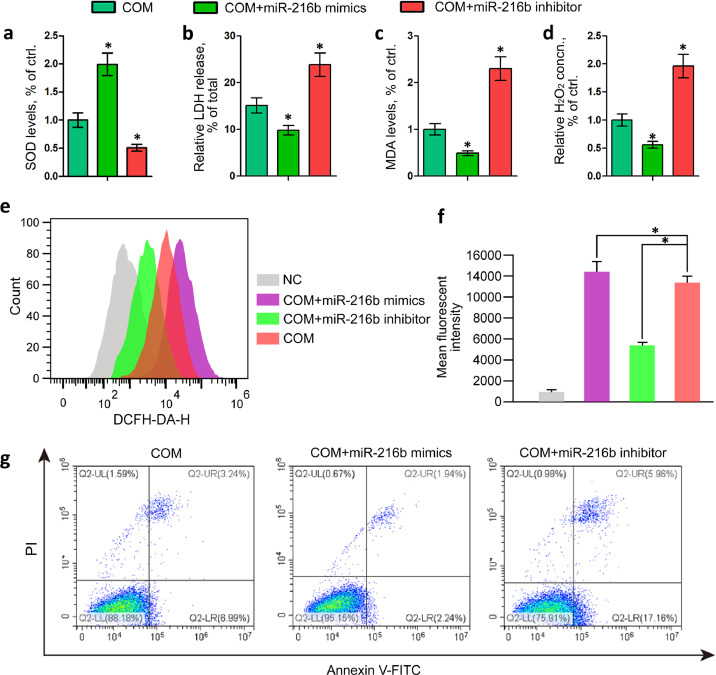
Background: Intrarenal calcium oxalate (CaOx) crystals induce inflammation and kidney tubular cell injury, which are processes that involve TLR4/NF-κB signalling. A recent genome-wide gene expression profile analysis of Randall's plaques in CaOx stone patients revealed that the expression of the long noncoding RNA H19 was significantly upregulated. However, to date, its role in kidney CaOx stones has not been reported.

Method: A Gene Expression Omnibus (GEO) dataset was utilized to analyse gene expression profiles. Luciferase reporter, Western blotting, qRT-PCR, immunofluorescence staining and reactive oxygen species (ROS) assays were employed to study the molecular mechanism of HMGB1/TLR4/NF-κB regulation by H19 and miR-216b. In vitro and in vivo assays were performed to further confirm the proinflammatory and prooxidative stress effects.

Finding: H19 expression was significantly increased and positively correlated with the expression levels of HMGB1, TLR4 and NF-κB in Randall's plaques and glyoxylate-induced CaOx nephrocalcinosis mouse models. H19 interacted with miR-216b and suppressed its expression. Additionally, miR-216b inhibited HMGB1 expression by directly binding its 3'-untranslated region. Moreover, H19 downregulation inhibited HMGB1, TLR4 and NF-κB expression and suppressed CaOx nephrocalcinosis-induced renal tubular epithelial cell injury, NADPH oxidase, and oxidative stress in vivo and in vitro. Interestingly, miR-216b inhibition partially reversed the inhibitory effect of H19 knockdown on HMGB1 expression.

Interpretation: We determined that H19 might serve as a facilitator in the process of CaOx nephrocalcinosis-induced oxidative stress and renal tubular epithelial cell injury, and we revealed that the interaction between H19 and miR-216b could exert its effect via the HMGB1/TLR4/NF-κB pathway.

Funding: This work was supported by the National Nature Science Foundation of China (Nos. 8196030190, 8190033175, 81370805, 81470935, 81900645, 81500534, and 81602236).

Keywords: H19; HMGB1; calcium oxalate; ceRNA; tubular epithelial cell injury.


Conflict of interest statement

The authors have nothing to disclose.

Figures

Fig 1
Fig. 1
H19 and HMGB1 expression was significantly increased in the CaOx nephrocalcinosis mouse model. (a) Hierarchical clustering and heatmap analysis of GSE73680 from a recent genome-wide gene expression profile analysis of Randall's plaques from 29 CaOx stone patients and 6 healthy controls. (b) A glyoxylate-induced kidney CaOx nephrocalcinosis mouse model was established and verified by polarized light microscopy (Magnification, × 100) and Pizzolato staining (Magnification, × 200) of CaOx crystal deposition in the corticomedullary junction area. PAS staining (Magnification, × 100) illustrating tubular injury. Immunohistochemical analysis of kidney HMGB1, TLR4 and NF-kB expression in a CaOx nephrocalcinosis-induced mouse model (Magnification, × 200). (c) qRT-PCR analysis was performed to detect the expression levels of HMGB1, TLR4 and NF-kB in CaOx mouse kidney samples, and these levels were compared with those in mock controls. Pearson correlation coefficient analysis of the expression levels of H19 and HMGB1 (d), TLR4 (e), and NF-kB (f). (g) qRT-PCR analysis was performed to detect the expression levels of HMGB1, TLR4 and NF-kB in HK-2 cells treated with different concentrations of COM crystals. The data are shown as the mean ± standard error (SE) of three independent experiments. (*P < 0.05; **P < 0.01, by Student's t-test (c) or Pearson's correlation test (d, e, f) or one-way ANOVA (g)).
Fig 2
Fig. 2
H19 facilitated CaOx nephrocalcinosis-induced renal tubular epithelial cell injury in vivo. CaOx deposition in the corticomedullary junction area was measured by polarized light microscopy (Magnification, × 100) and Pizzolato staining (Magnification, × 200). PAS staining (Magnification, × 100) illustrating renal tubular epithelial cell injury. (b) Immunohistochemical analysis of kidney HMGB1, TLR4, NF-kB, SOD2 and NOX2 expression was performed in rAAV-H19 or rAAV-vector injected CaOx nephrocalcinosis mouse models (Magnification, × 200). (c) Western blot analysis was performed to detect HMGB1, TLR4, p-NF-kB and NF-kB protein expression in H19-treated kidney tissue. GAPDH served as an internal control. (d) qRT-PCR analysis was performed to detect the expression levels of proinflammatory cytokines in kidney tissue. The data are shown as the mean ± standard deviation (SD) of three independent experiments. (*P < 0.05; **P < 0.01, by Student's t-test (a, b, d)).
Fig 3
Fig. 3
H19 promoted COM crystal-induced renal tubular epithelial cell oxidative stress injury in vitro. Western blot (a) and qRT-PCR (b) analyses of HMGB1, TLR4, NF-kB and p-NF-kB expression in HK-2 cells. β-Actin was used for normalization. SOD level (c), LDH release (d), MDA level (e), and H2O2 concentration (f) were determined in HK-2 cells incubated with COM crystals following lenti-H19 and si-H19 treatment. (g) Cellular ROS production in HK-2 cells was measured by flow cytometry. (h) Histograms showing the mean fluorescence intensity of DCFH. (i) The apoptosis effect was investigated by flow cytometric analysis of HK-2 cells stained with Annexin V-FITC and propidium iodide. (*P < 0.05; **P < 0.01, by one-way ANOVA (b-f, h, i)).
Fig 4
Fig. 4
H19 interacted with miR-216b by directly binding its 3′-UTR. Schematic diagram of the mutant and WT seed sequences of miR-216b targeting the 3′-UTR of H19. (b) qRT-PCR analysis was performed to detect the expression level of miR-216b in CaOx-induced mouse kidney samples, and this expression was compared with that in the mock control. U6 was used as a miRNA control. (c) Pearson correlation coefficient analysis of the expression levels of H19 and miR-216b. (d) miR-216b expression was inhibited by lenti-H19 transfection. (e) H19 expression was detected using qRT-PCR in HK-2 cells transfected with the miR-216b mimics and inhibitor. Luciferase reporters harbouring putative target sites in the WT and mutant 3′-UTR of H19 were cotransfected with 100 nM miR-216b mimics (f) or miR-216b inhibitor (g) in HK-2 cells. The data are shown as the mean ± SD of three independent experiments. (*P < 0.05; **P < 0.01, by Student's t-test (b) or Pearson's correlation test (c) or one-way ANOVA (d-g)).
Fig 5
Fig. 5
miR-216b inhibited HMGB1 expression by directly binding to its 3′-UTR. Schematic diagram of our constructed luciferase reporter plasmid psiCHECK-2 containing the 3′-UTR of HMGB1. (b) Mutant and WT seed sequences of miR-216b targeting the 3′-UTR of HMGB1. (c) Luciferase reporters harbouring putative target sites in the WT and mutant 3′-UTRs of HMGB1 were cotransfected with 100 nM of the indicated small RNA molecules in HK-2 cells. (d) Pearson correlation coefficient analysis of the expression levels of H19 and miR-216b. (e) Western blot analysis was performed to detect the expression of HMGB1, TLR4, NF-kB and p-NF-kB in HK-2 cells transfected with the miR-216b mimics or inhibitor. Actin-β served as an internal control. (f) miR-216b mimics and inhibitor were used to establish miR-216b overexpression and inhibition, respectively, in HK-2 cells. The expression of HMGB1 (g), TLR4 (h), and NF-kB (i) was detected using qRT-PCR in HK-2 cells transfected with miR-216b mimics or inhibitor. The data are shown as the mean ± SD of three independent experiments. (*P < 0.05; **P < 0.01, by one-way ANOVA (c, e-i) or Pearson's correlation test (d)).
Fig 6
Fig. 6
miR-216b suppressed COM crystal-induced renal tubular epithelial cell oxidative stress injury in vitro. (a) SOD level, (b) LDH release, (c) MDA level, and (d) H2O2 concentration were determined in HK-2 cells incubated with COM crystals following miR-216b mimics or inhibitor treatment. (e-f) Cellular ROS production in HK-2 cells was measured by flow cytometry. Histograms showing the mean fluorescence intensity of DCFH. (g) Apoptosis was analysed by flow cytometry in HK-2 cells transfected with miR-216b mimics or inhibitor. Histograms showing the mean fluorescence intensity. The data are shown as the mean ± SD of three independent experiments. (*P < 0.05; **P < 0.01, by one-way ANOVA (a-d, f, g)).
Fig 7
Fig. 7
miR-216b reversed the effect of H19 on CaOx nephrocalcinosis-induced renal tubular epithelial cell injury in vivo. (a) CaOx deposition in the corticomedullary junction area was measured by polarized light microscopy (Magnification, × 100) and Pizzolato staining (Magnification, × 200). PAS staining (Magnification, × 100) illustrating renal tubular epithelial cell injury. (b) Immunohistochemical analysis of kidney HMGB1, TLR4, NF-kB, SOD2 and NOX2 expression was performed in rAAV-H19-, miR-216b agonist-, or combination-treated CaOx nephrocalcinosis mouse models (magnification in all panels is 200 ×). Quantifications were performed using ImageJ. The data are shown as the mean ± SD of three independent experiments. (*P < 0.05; **P < 0.01, by one-way ANOVA (a, b)).
Fig 8
Fig. 8
miR-216b reversed the effect of H19 on COM crystal-induced renal tubular epithelial cell oxidative stress injury in vitro. Western blot (a) and qRT-PCR (b) analyses of HMGB1, TLR4 and NF-kB expression in HK-2 cells following transfection with lenti-H19, miR-216b mimics, or their combination. β-Actin was used for normalization. ROS generation (c), LDH release (d), cellular malondialdehyde (MDA) levels (e), and H2O2 concentrations (f) were determined in HK-2 cells incubated with COM crystals following treatment with lenti-H19, miR-216b mimics or their combination. (G-H) Flow cytometric analysis of cellular ROS generation was carried out in HK-2 cells. Histograms showing the mean fluorescence intensity of DCFH. (g) The apoptosis effect was investigated by flow cytometric analysis of HK-2 cells stained with Annexin V-FITC and propidium iodide. The data are shown as the mean ± SD of three independent experiments. (*P < 0.05; **P < 0.01, by one-way ANOVA (b–i)).
Fig 9
Fig. 9
Schematic model. LncRNA H19 sponges miR-216b-3p to promote calcium oxalate nephrocalcinosis-induced renal tubular epithelial cell injury via HMGB1/TLR4/NF-κB pathway activation.

References

    1. Khan S.R., Pearle M.S., Robertson W.G. Kidney stones. Nat Rev Dis Primers. 2016;2:16008. - PMC - PubMed
    1. Zeng G., Mai Z., Xia S. Prevalence of kidney stones in China: an ultrasonography based cross-sectional study. BJU Int. 2017;120(1):109–116. - PubMed
    1. Mulay S.R., Eberhard J.N., Desai J. Hyperoxaluria requires tnf receptors to initiate crystal adhesion and kidney stone disease. J Am Soc Nephrol. 2017;28(3):761–768. - PMC - PubMed
    1. Ruan Y., Wang L., Zhao Y. Carbon monoxide potently prevents ischemia-induced high-mobility group box 1 translocation and release and protects against lethal renal ischemia-reperfusion injury. Kidney Int. 2014;86(3):525–537. - PubMed
    1. Chen X., Wu S., Chen C. Omega-3 polyunsaturated fatty acid supplementation attenuates microglial-induced inflammation by inhibiting the HMGB1/TLR4/NF-kappaB pathway following experimental traumatic brain injury. J Neuroinflammation. 2017;14(1):143. - PMC - PubMed

MeSH terms