. 2019 Dec 27;294(52):19844-19851.
doi: 10.1074/jbc.AC119.010611. Epub 2019 Nov 17.

Interferon-induced transmembrane proteins inhibit cell fusion mediated by trophoblast syncytins

Affiliations

Interferon-induced transmembrane proteins inhibit cell fusion mediated by trophoblast syncytins

Ashley Zani et al. J Biol Chem. .

Abstract

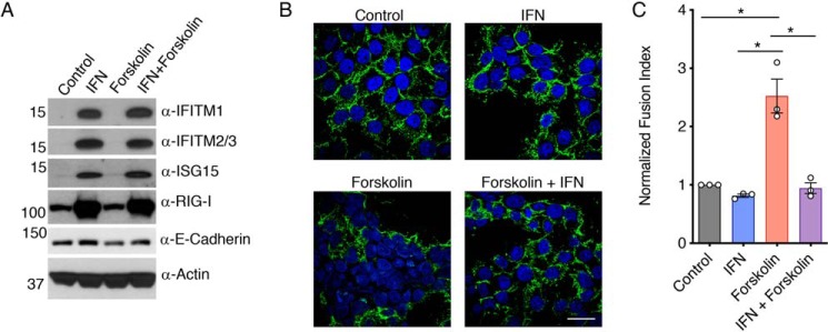
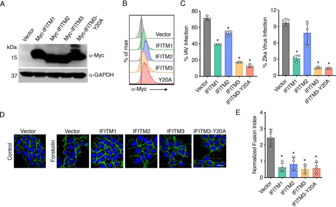
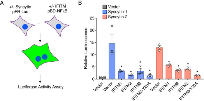
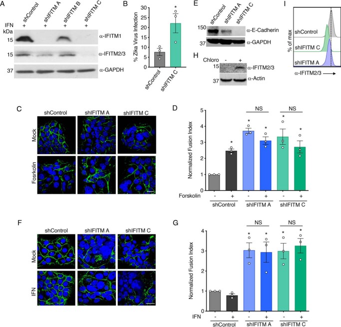
Type I interferon (IFN) induced by virus infections during pregnancy can cause placental damage, but the mechanisms and identities of IFN-stimulated genes that are involved in this damage remain under investigation. The IFN-induced transmembrane proteins (IFITMs) inhibit virus infections by preventing virus membrane fusion with cells and by inhibiting fusion of infected cells (syncytialization). Fusion of placental trophoblasts via expression of endogenous retroviral fusogens known as syncytins forms the syncytiotrophoblast, a multinucleated cell structure essential for fetal development. We found here that IFN blocks fusion of BeWo human placental trophoblasts. Stably expressed IFITM1, -2, and -3 also blocked fusion of these trophoblasts while making them more resistant to virus infections. Conversely, stable IFITM knockdowns in BeWo trophoblasts increased their spontaneous fusion and allowed fusion in the presence of IFN while also making the cells more susceptible to virus infection. We additionally found that exogenous expression of IFITMs in HEK293T cells blocked fusion with cells expressing syncytin-1 or syncytin-2, confirming the ability of IFITMs to block individual syncytin-mediated fusion. Overall, our data indicate that IFITMs inhibit trophoblast fusion and suggest that there may be a critical balance between these antifusogenic effects and the beneficial antiviral effects of IFITMs in virus infections during pregnancy.

Keywords: IFITM; IFITM1; IFITM2; IFITM3; Zika virus; cell fusion; fusion protein; influenza virus; interferon; interferon-induced transmembrane protein; membrane fusion; placenta; restriction factor; syncytin; trophoblast; virus.


Conflict of interest statement

The authors declare that they have no conflicts of interest with the contents of this article

Figures

Figure 1.
Figure 1.
IFN inhibits fusion of BeWo trophoblasts stimulated by forskolin. BeWo cells were treated for 24 h with 40 units/ml IFNβ (IFN), 50 μm forskolin, a combination of IFN and forskolin, or vehicle control. Treated cells were analyzed by Western blotting of cell lysates (A) or confocal microscopy imaging (B) after staining with anti-E-cadherin (green) or DAPI (blue). White scale bar, 10 μm. C, fusion indices were calculated and normalized to the background spontaneous fusion level observed in each experiment in vehicle control cells. Bars, averages from three identical independent experiments with individual data points shown as circles; data are representative of two additional similar experiments. Error bars, S.D. Differences between groups were evaluated by one-way ANOVA followed by Tukey's multiple-comparison test. *, p < 0.0005 for the indicated comparisons.
Figure 2.
Figure 2.
IFITM overexpression inhibits BeWo trophoblast fusion. BeWo cells were stably transduced with lentiviruses expressing Myc-tagged IFITM constructs. A, Western blotting of cell lysates. B, flow cytometry histograms after staining with anti-Myc antibody. C, percentage of infection measured 24 h postinfection with influenza A virus (IAV) (MOI 2.5) or Zika virus (MOI 5), as measured by flow cytometry staining for viral antigens. Flow cytometry gates were set based on mock-infected controls. Bars, averages of three independent experiments with individual data points shown as circles. Error bars, S.D. Differences between groups were analyzed by one-way ANOVA followed by Dunnett's multiple-comparison test. *, p < 0.0001 compared with vector control cells. D, cell lines were treated for 48 h with 50 μm forskolin or vehicle control. Cells were then imaged by confocal microscopy after staining with anti-E-cadherin (green) or DAPI (blue). White scale bar, 10 μm. E, fusion indices from forskolin-treated cells as in D were calculated and normalized to the background spontaneous fusion levels observed in each experiment in vector control cells treated with vehicle control. Bars, averages from four independent experiments with individual data points shown as circles. Error bars, standard S.D. Differences between groups were analyzed by one-way ANOVA followed by Dunnett's multiple-comparison test. *, p < 0.0001 compared with vector control cells.
Figure 3.
Figure 3.
IFITM inhibition of syncytin-mediated fusion of HEK293T cells. A, schematic representation of the HEK293T cell fusion assay. One population of cells was transfected with syncytin-1 or -2 constructs or vector control plus pFR-Luc plasmid. A second population of cells was transfected with IFITM constructs or vector control plus pBD-NF-κB. Cells were then mixed and allowed to fuse for 24 h. Luciferase activity in cell lysates was then measured as a quantitative readout of cell fusion. B, luciferase activity was measured from cells prepared as described in A. Bars, average results from three independent experiments with individual data points shown as circles. Error bars, S.D. Differences between groups were analyzed by one-way ANOVA followed by Tukey's multiple-comparison test. *, p < 0.01 compared with the respective syncytin-transfected vector control cells.
Figure 4.
Figure 4.
Knockdown of IFITMs promotes fusion of BeWo trophoblasts. BeWo cells were stably transduced with lentiviruses expressing control shRNA (shControl) or different shRNAs targeting IFITMs (shIFITM A, B, or C). A, Western blotting of cell lysates after 18-h treatment with 40 units/ml IFNβ demonstrates IFITM knockdown in specific cell lines. B, percentage of infection of shControl cells and shIFITM C cells 24 h postinfection with Zika virus (MOI 5), as measured by flow cytometry staining for viral antigen. Flow cytometry gates were set based on mock-infected controls. Bars, averages of three independent experiments with individual data points shown as circles. Error bars, S.D. *, p < 0.05 as compared with shControl cells by paired t test. C, the indicated cell lines were treated for 48 h with 50 μm forskolin or vehicle control. Cells were then imaged by confocal microscopy after staining with anti-E-cadherin (green) or DAPI (blue). White scale bar, 10 μm. D, fusion indices from cells as in C were calculated and normalized to the background spontaneous fusion levels observed in each experiment in shControl cells treated with vehicle control. Bars, averages from three independent experiments with individual data points shown as circles. Error bars, S.D. Differences between groups were evaluated by one-way ANOVA followed by Tukey's multiple-comparison test. *, p < 0.02 compared with shControl without forskolin treatment. NS, selected comparisons of interest that are not significantly different as judged by a p value > 0.05. E, Western blotting of cell lysates demonstrates down-regulation of E-cadherin in IFITM knockdown lines. F, the indicated cell lines were treated for 48 h with 40 units/ml IFNβ or vehicle control. Cells were then imaged by confocal microscopy after staining with anti-E-cadherin (green) or DAPI (blue). White scale bar, 10 μm. G, fusion indices from cells as in F were calculated and normalized to the background spontaneous fusion levels observed in each experiment in shControl cells treated with vehicle control. Bars, averages from three independent experiments with individual data points shown as circles. Error bars, S.D. Differences between groups were evaluated by one-way ANOVA followed by Tukey's multiple-comparison test. *, p < 0.01 compared with shControl without IFNβ treatment. NS, selected comparisons of interest that are not significantly different as judged by a p value > 0.05. H, Western blotting of WT BeWo cells after 24-h treatment with 40 μm chloroquine (chloro) or vehicle control (mock) demonstrates accumulation of baseline IFITM2/3 when lysosomal degradation is inhibited. I, flow cytometry histograms for untreated/unstimulated cell lines after staining with anti-IFITM2/3 antibody confirm that low baseline levels of IFITMs are reduced by shRNAs targeting IFITMs.

References

    1. Coyne C. B., and Lazear H. M. (2016) Zika virus: reigniting the TORCH. Nat. Rev. Microbiol. 14, 707–715 10.1038/nrmicro.2016.125 - DOI - PubMed
    1. Hirsch A. J., Roberts V. H. J., Grigsby P. L., Haese N., Schabel M. C., Wang X., Lo J. O., Liu Z., Kroenke C. D., Smith J. L., Kelleher M., Broeckel R., Kreklywich C. N., Parkins C. J., Denton M., et al. (2018) Zika virus infection in pregnant rhesus macaques causes placental dysfunction and immunopathology. Nat. Commun. 9, 263 10.1038/s41467-017-02499-9 - DOI - PMC - PubMed
    1. Platt D. J., Smith A. M., Arora N., Diamond M. S., Coyne C. B., and Miner J. J. (2018) Zika virus-related neurotropic flaviviruses infect human placental explants and cause fetal demise in mice. Sci. Transl. Med. 10, eaao7090 10.1126/scitranslmed.aao7090 - DOI - PMC - PubMed
    1. Yockey L. J., Jurado K. A., Arora N., Millet A., Rakib T., Milano K. M., Hastings A. K., Fikrig E., Kong Y., Horvath T. L., Weatherbee S., Kliman H. J., Coyne C. B., and Iwasaki A. (2018) Type I interferons instigate fetal demise after Zika virus infection. Sci. Immunol. 3, eaao1680 10.1126/sciimmunol.aao1680 - DOI - PMC - PubMed
    1. Casazza R. L., and Lazear H. M. (2018) Antiviral immunity backfires: pathogenic effects of type I interferon signaling in fetal development. Sci. Immunol. 3, eaar3446 10.1126/sciimmunol.aar3446 - DOI - PubMed

Publication types

MeSH terms

LinkOut - more resources