. 2020 Sep-Oct;22(5):493-499.
doi: 10.4103/aja.aja_122_19.

Phosphoribosyl-pyrophosphate synthetase 2 (PRPS2) depletion regulates spermatogenic cell apoptosis and is correlated with hypospermatogenesis

Affiliations

Phosphoribosyl-pyrophosphate synthetase 2 (PRPS2) depletion regulates spermatogenic cell apoptosis and is correlated with hypospermatogenesis

Bin Lei et al. Asian J Androl. 2020 Sep-Oct.

Abstract

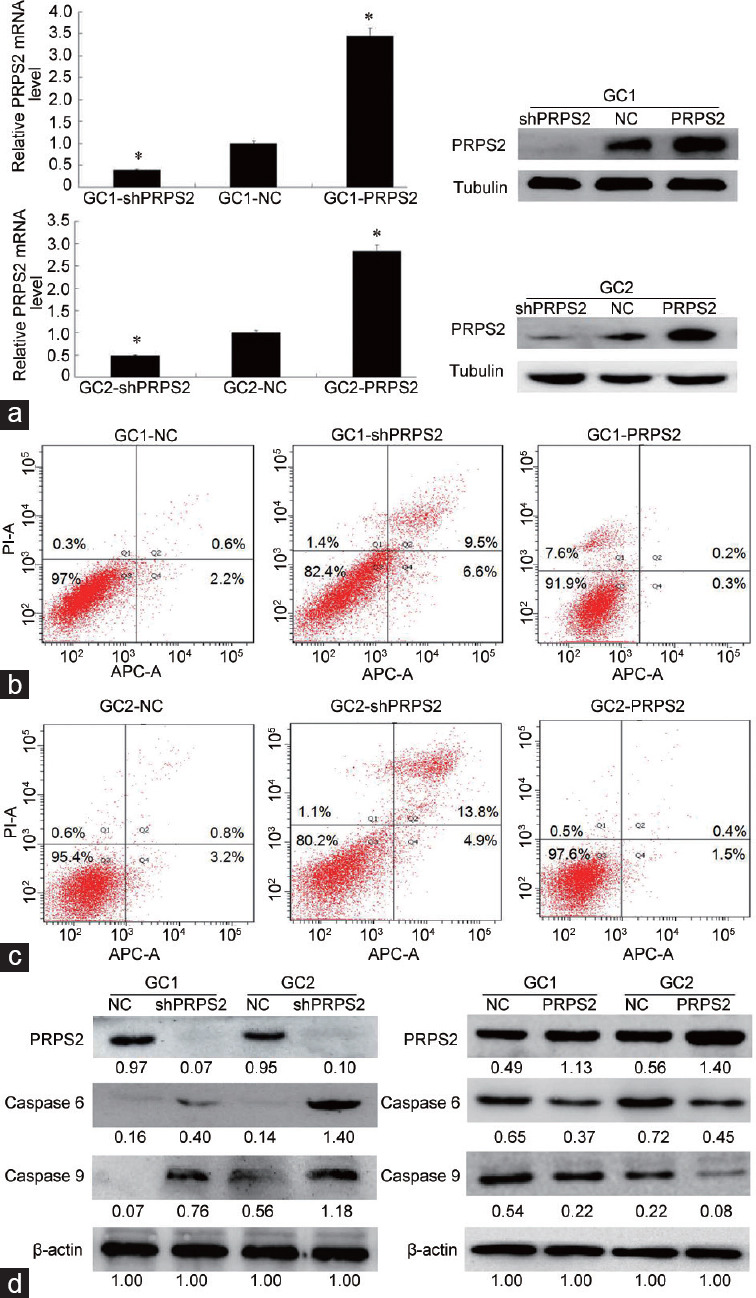
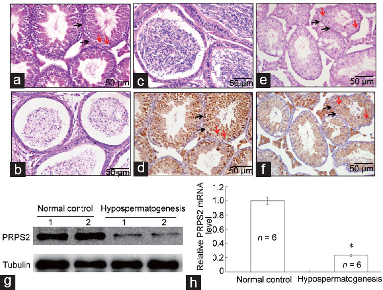
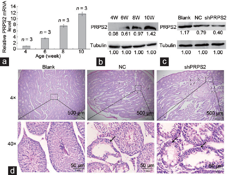
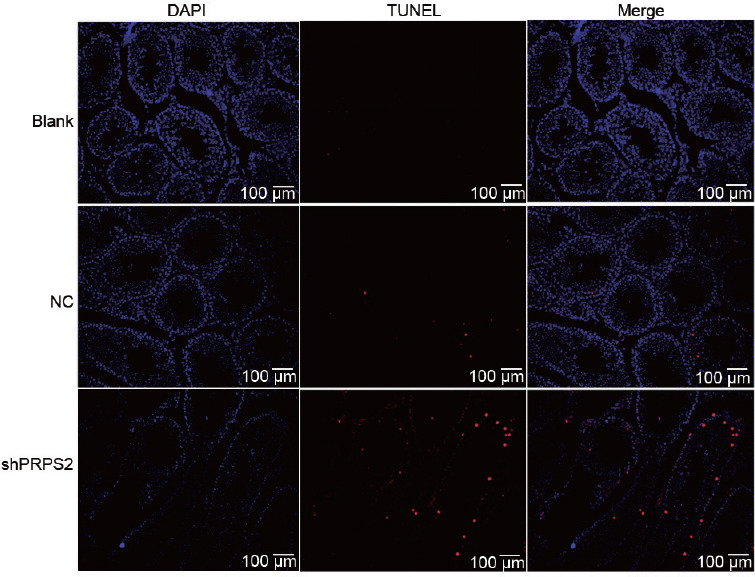
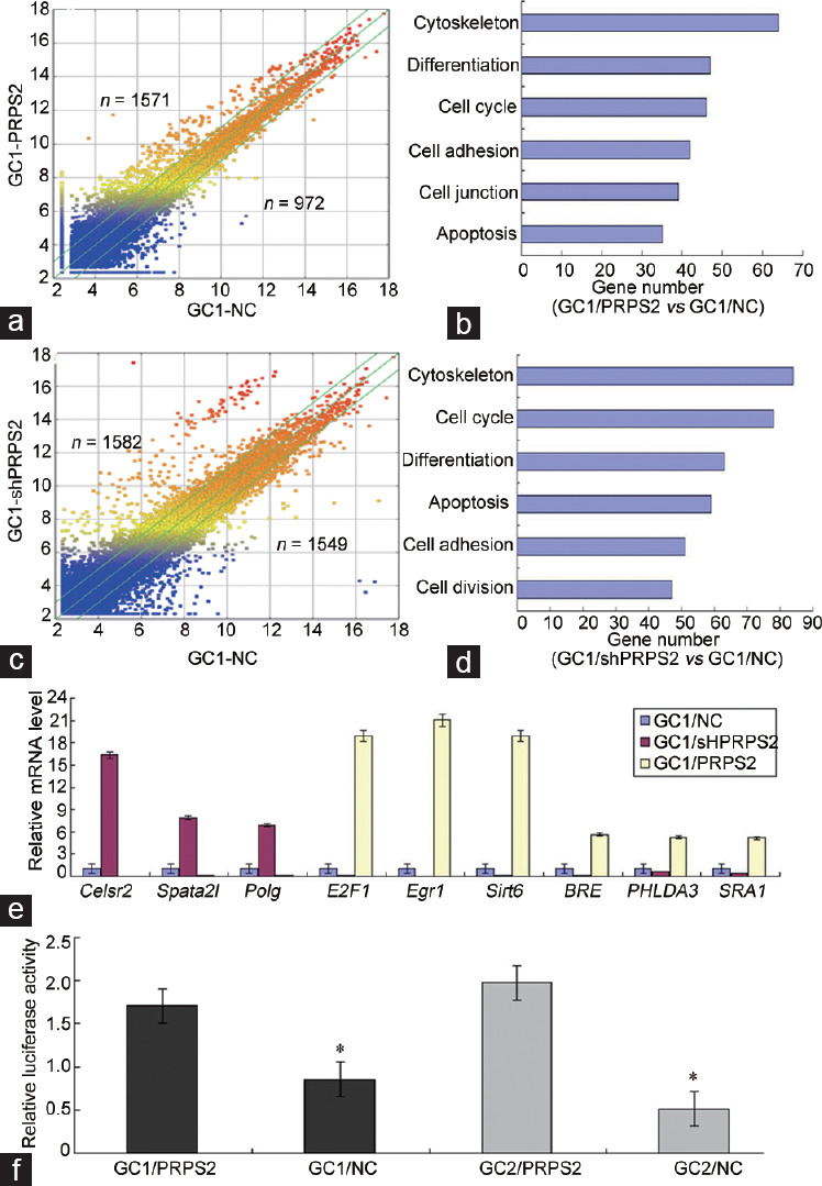
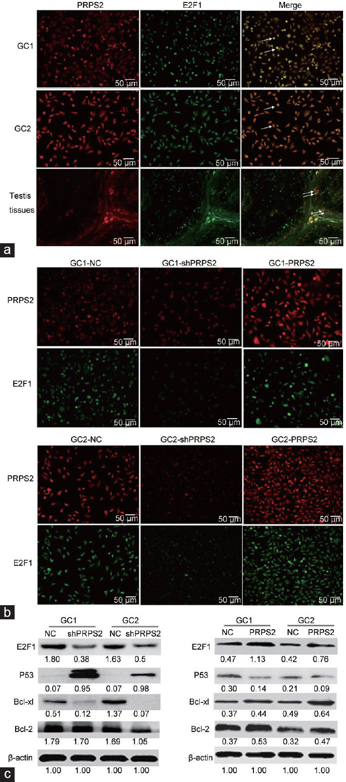
Phosphoribosyl-pyrophosphate synthetase 2 (PRPS2) is a rate-limiting enzyme and plays an important role in purine and pyrimidine nucleotide synthesis. Recent studies report that PRPS2 is involved in male infertility. However, the role of PRPS2 in hypospermatogenesis is unknown. In this study, the relationship of PRPS2 with hypospermatogenesis and spermatogenic cell apoptosis was investigated. The results showed that PRPS2 depletion increased the number of apoptotic spermatogenic cells in vitro. PRPS2 was downregulated in a mouse model of hypospermatogenesis. When PRPS2 expression was knocked down in mouse testes, hypospermatogenesis and accelerated apoptosis of spermatogenic cells were noted. E2F transcription factor 1 (E2F1) was confirmed as the target gene of PRPS2 and played a key role in cell apoptosis by regulating the P53/Bcl-xl/Bcl-2/Caspase 6/Caspase 9 apoptosis pathway. Therefore, these data indicate that PRPS2 depletion contributes to the apoptosis of spermatogenic cells and is associated with hypospermatogenesis, which may be helpful for the diagnosis of male infertility.

Keywords: hypospermatogenesis; male infertility; molecular marker; phosphoribosyl-pyrophosphate synthetase 2; spermatogenesis.


Conflict of interest statement

None

Figures

Figure 1
Figure 1
PRPS2 depletion and apoptosis of spermatogenic cells. (a) qRT-PCR and Western blot analysis confirmed that PRPS2 was successfully downregulated by gene silencing and upregulated by PRPRS2 overexpression in both GC1 and GC2 cells. (b) Flow cytometry analysis shows that PRPS2 depletion promotes the apoptosis of GC1 cells. (c) Flow cytometry analysis shows that PRPS2 depletion promotes the apoptosis of GC2 cells (abscissa: cell count; ordinate: the fluorescence intensity). (d) Western blot analysis indicates that PRPS2 depletion activates the expression of apoptotic proteins (values beneath blots are relative to the control). *Significantly different compared with negative control, P < 0.05 by independent-samples t-test. shPRPS2: small-hairpin RNA gene silencer; NC: negative control; PRPS2: overexpression; PRPS2: phosphoribosyl-pyrophosphate synthetase 2; qRT-PCR: quantitative real-time polymerase chain reaction; APC-A: Allophycocyanin-A.
Figure 2
Figure 2
Correlation of PRPS2 expression with hypospermatogenesis. (a) H and E staining of the testis with normal spermatogenesis. (b) H and E staining of normal cauda epididymidis. (c) H and E staining of a testis with hypospermatogenesis. (d) H and E staining of cauda epididymidis in hypospermatogenesis. (e) PRPS2 expression was detected in normal testis by IHC. (f) PRPS2 expression was detected in a testis with hypospermatogenesis by IHC. Scale bars = 50 μm. (g) PRPS2 expression was detected in testis with normal spermatogenesis and hypospermatogenesis by Western blot. (h) PRPS2 expression was detected in testis tissues with normal spermatogenesis and hypospermatogenesis by qRT-PCR. *Hypospermatogenesis group compared with normal control, P < 0.05 by independent-samples t-test. Red arrow: spermatogonia; Black arrow: spermatocyte. PRPS2: phosphoribosyl-pyrophosphate synthetase 2; H and E: hematoxylinand and eosin; IHC: immunohistochemical; qRT-PCR: quantitative real-time polymerase chain reaction.
Figure 3
Figure 3
PRPS2 depletion and hypospermatogenesis. (a) PRPS2 mRNA levels were increased with age in the testis. (b) PRPS2 protein level increased with age in the testis. (c) When treated with murine PRPS2-specific shRNA lentivirus, PRPS2 downregulation was observed in testis tissues at 9 weeks of age. (d) When treated with murine PRPS2-specific shRNA lentivirus, hypospermatogenesis was observed in the testis. Upper row: low-power magnification, scale bars=500 μm. Lower row: high-power magnification, scale bars=50 μm. Black arrow: damaged seminiferous tubules. PRPS2: phosphoribosyl-pyrophosphate synthetase 2; NC: negative control; W: weeks.
Figure 4
Figure 4
Apoptotic cell number visualized by the TUNEL assay. Representative micrographs indicating an increase in number after treatment with murine PRPS2-specific shRNA lentivirus. PRPS2: phosphoribosyl-pyrophosphate synthetase 2; TUNEL: transferase-mediated deoxyuridine triphosphate-biotin nick end labeling; DAPI: 4',6-diamidino-2-phenylindole.
Figure 5
Figure 5
The potential genes and pathways regulated by PRPS2 were identified in GC1 cells. (a) Scatter diagram of the differential genes in GC1/PRPS2 and GC1/NC cells. (b) Potential signaling pathways of the differential genes in GC1/PRPS2 and GC1/NC cells. (c) Scatter diagram of the differential genes in GC1/shPRPS2 and GC1/NC cells. (d) Potential signaling pathways of the differential genes in GC1/shPRPS2 and GC1/NC cells. (e) Nine genes associated with cell apoptosis signal were confirmed in GC1 cells by qRT-PCR. (f) Transcriptional activity of E2F1 was measured by Luciferase Reporter Assay. *GC1/NC versus GC1/PRPS2 and GC2/NC versus GC2/PRPS2, P < 0.05 by independent-samples t-test. PRPS2: phosphoribosyl-pyrophosphate synthetase 2; NC: negative control; qRT-PCR: quantitative real-time polymerase chain reaction; Polg: polymerase (DNA directed), gamma; E2F1: E2F transcription factor 1; Egr1: early growth response 1; BRE: brain and reproductive organ-expressed; PHLDA3: pleckstrin homology-like domain family A member 3; SRA1: steroid receptor RNA activator 1.
Figure 6
Figure 6
PRPS2 targetes E2F1 and activation of the P53/Bcl-xl/Bcl-2 signal pathway. (a) Colocation of PRPS2 and E2F1 in GC1 and GC2 cells and normal testis tissue was observed by double immunofluorescent staining. (b) After transfection, the expression levels of PRPS2 and E2F1 were detected by double immunofluorescent staining. (c) Western blot was used to measure the expression levels of the E2F1/P53/Bcl-xl/Bcl-2 signaling pathway. Values beneath the blots are relative to the control. White arrow: colocation. PRPS2: phosphoribosyl-pyrophosphate synthetase 2; NC: negative control; E2F1: E2F transcription factor 1.

Similar articles

Cited by

References

    1. Robin G, Boitrelle F, Leroy X, Peers MC, Marcelli F, et al. Assessment of azoospermia and histological evaluation of spermatogenesis. Ann Pathol. 2010;30:182–95. - PubMed
    1. Vogelweid CM, Verina T, Norton J, Harruff R, Ghetti B. Hypospermatogenesis is the cause of infertility in the male weaver mutant mouse. J Neurogenet. 1993;9:89–104. - PubMed
    1. Sugimoto K, Koh E, Iijima M, Taya M, Maeda Y, et al. Aberrant methylation of the TDMR of the GTF2A1L promoter does not affect fertilisation rates via TESE in patients with hypospermatogenesis. Asian J Androl. 2013;15:634–9. - PMC - PubMed
    1. Takeshima T, Yumura Y, Iwasaki A, Noguchi K. Clinical review of hypospermatogenesis in patients with a previous episode of mumps orchitis. Hinyokika Kiyo. 2015;61:227–33. Article in Japanese. - PubMed
    1. Almeida C, Correi S, Rocha E, Alves A, Ferraz L, et al. Caspase signalling pathways in human spermatogenesis. J Assist Reprod Genet. 2013;30:487–95. - PMC - PubMed

Publication types

MeSH terms