. 2018 Oct;11(3):189-199.
doi: 10.2478/intox-2018-0016. Epub 2019 Oct 18.

Insights on the relationship between structure vs. toxicological activity of antibacterial rhodamine-labelled 3-hydroxy-4-pyridinone iron(III) chelators in HepG2 cells

Affiliations

Insights on the relationship between structure vs. toxicological activity of antibacterial rhodamine-labelled 3-hydroxy-4-pyridinone iron(III) chelators in HepG2 cells

Tânia Moniz et al. Interdiscip Toxicol. 2018 Oct.

Abstract

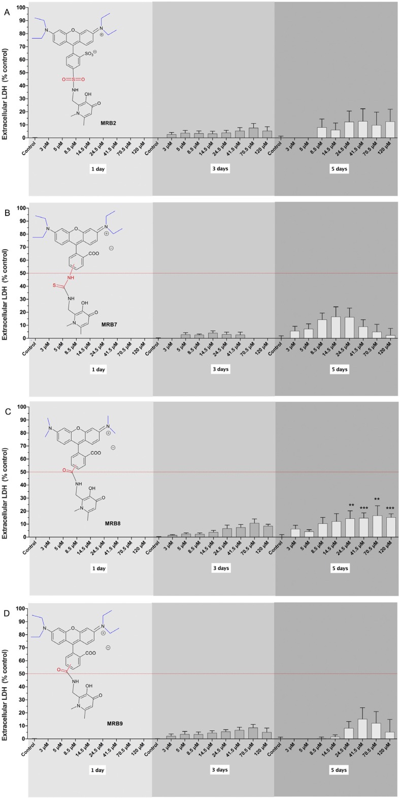
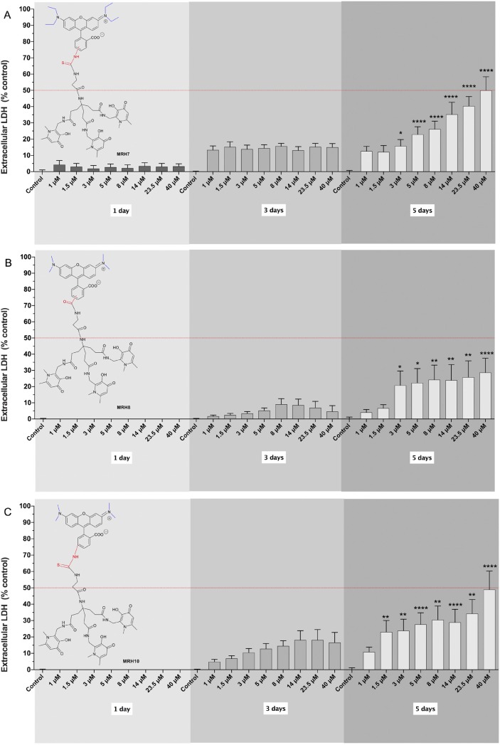
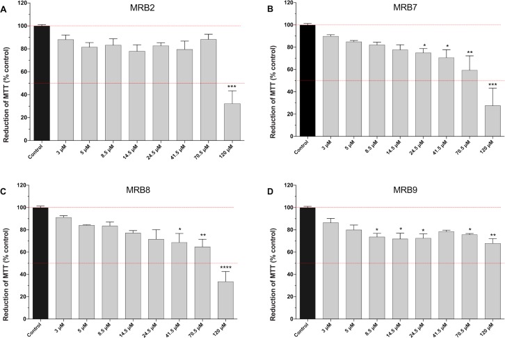
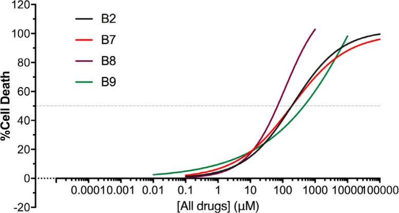
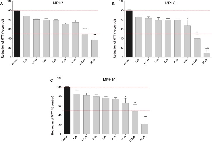
In the present study we investigated the in vitro hepatotoxicity of a set of rhodamine-labelled 3-hydroxy-4-pyridinones (3,4-HPO) that had previously demonstrated significant inhibitory effect in the intramacrophagic growth of Mycobacterium avium. Our aim was to establish a correspondence between the molecular structure and the in vitro toxicological activity of these compounds. The impact of a set of bidentate (MRB2, MRB7, MRB8, and MRB9) and hexadentate (MRH7, MRH8, and MRH10) chelators on cellular metabolic competence and membrane integrity was investigated in HepG2 cells. Our findings indicate that: a) hexadentate chelators are more cytotoxic than parent bidentate ligands; b) disruption of cell membrane and metabolic competence only occurred after 5 days, at the highest concentrations tested; c) strict correlation between bacteriostatic activity and in vitro toxicity was observed, which seems to be directly dependent on the size of the molecule and on the hydrophilic/lipophilic balance; d) among the set of bidentate ligands, carboxyrhodamine derivatives (amide linker) presented lower detrimental effects, when compared with rhodamine B isothiocyanate chelators (thiourea linker); e) contrarily, for the hexadentate series, rhodamine B isothiocyanate derivatives are less cytotoxic to HepG2 cells than carboxyrhodamine molecules; and f) for all compounds tested, when the substituents of the nitrogen atom were switched from ethyl to methyl, an increment of toxicity was observed. Overall, all chelators seem to display suitable in vitro toxicological potential to combat fast grow bacteria. According to their in vitro pharmacological: toxicological potential ratio, MRH7 and MRH8 may be considered as the most suitable compounds to undergo further pre-clinical development studies.

Keywords: 3-hydroxy-4-pyridinone (3,4-HPO); HepG2 cells; in vitrotoxicity; iron chelator; rhodamine.


Figures

Figure 1
Figure 1
Structure and nomenclature of fluorescent bidentate (MRB2, MRB7, MRB8, and MRB9) and hexadentate (MRH7, MRH8, and MRH10) chelators used in the current study. In previous works, the compound MRH7 was abbreviated as CP777 (Fernandes et al., 2010) and 4 (Nunes et al., 2010). The molecular structure of these chelators comprise three parts: i) a chelating unit, i.e. 3-hydroxy-4-pyridinone (3,4-HPO); ii) a fluorophore (tetraethyl sulphorhodamine B for MRB2; tetraethyl rhodamine B isothiocyanate for MRB7 and MRH7; tetramethyl carboxyrhodamine for MRB8 and MRH8; tetraethyl carboxyrhodamine for MRB9; or tetramethyl rhodamine B isothiocyanate for MRH10); and iii) a linkage fragment, which is determined by the fluorophore derivative that is used in the synthesis. The chelating unit is directly coupled to the fluorophore to produce the bidentate fluorescent ligands (MRB2, MRB7, MRB8, and MRB9) or linked to a tripodal anchor to produce hexadentate chelators (MRH7, MRH8, and MRH10). The substituents on xanthene group of rhodamine are highlighted in blue. The linker between the rhodamine fluorophore and the chelating unit is highlighted in red.
Figure 2
Figure 2
Cell membrane disruption assessed by the leakage of lactate dehydrogenase (LDH) to the extracellular medium, in Hep G2 cells, following exposure to bidentate chelators, i.e. MRB2 (A), MRB7 (B), MRB8 (C), and MRB9 (D), for 1, 3, or 5 days, at 37 °C. Each chelator was tested at concentrations ranging from 3 to 120 μM. Data are presented as mean ± standard error of the mean (SEM) of percentage of extracellular LDH, relative to the positive controls (cells treated with 1% Triton-X100), and were obtained in four independent experiments, performed in triplicates. The dashed red line represents half of the percentage of LDH leakage obtained for the positive controls (50%). Statistical analysis was performed using Kruskal-Wallis test followed by Dunn’s multiple comparison post hoc test. *p<0.05; **p<0.01 and ***p<0.001, when compared with controls at the same time-point.
Figure 3
Figure 3
Cell membrane disruption assessed by the leakage of lactate dehydrogenase (LDH) to the extracellular medium, in Hep G2 cells, following exposure to hexadentate chelators, i.e. MRH7 (A), MRH8 (B), and MRH10 (C), for 1, 3, or 5 days, at 37 °C. Each chelator was tested at concentrations ranging from 1 to 40 μM. Data are presented as mean ± standard error of the mean (SEM) of percentage of extracellular LDH, relative to the positive controls (cells treated with 1% Triton-X100), and were obtained in four independent experiments, performed in triplicates. The dashed red line represents half of the percentage of LDH leakage obtained for the positive controls (50%). Statistical analysis was performed using Kruskal-Wallis test followed by Dunn’s multiple comparison post hoc test. *p<0.05, **p<0.01 and ***p<0.001, when compared with controls at the same time-point.
Figure 4
Figure 4
Cell metabolic competence assessed by the reduction of the MTT assay, following exposure of HepG2 cells to bidentate chelators, i.e. MRB2 (A), MRB7 (B), MRB8 (C), and MRB9 (D), for 5 days, at 37 °C. Each chelator was tested at concentrations ranging from 3 to 120 μM. Data are presented as mean ± standard error of the mean (SEM) of percentage of MTT reduction, relative to the negative controls (black bar), and were obtained in four independent experiments, performed in triplicates. The dotted red lines represent 50% and 100% effect. Statistical analysis was performed using Kruskal-Wallis test followed by Dunn’s multiple comparison post hoc test. *p<0.05, **p<0.01, and *** or **** p<0.001, when compared with control.
Figure 5
Figure 5
Regression models for the cell metabolic competence, as assessed by the reduction of the MTT assay, following exposure of HepG2 cells to bidentate chelators, i.e. MRB2 (black line), MRB7 (red line), MRB8 (purple line), and MRB9 (green line), for 5 days, at 37 °C. Data relative to the negative controls were from four independent experiments run in triplicate. The dotted line represent 50% effect.
Figure 6
Figure 6
Cell metabolic competence, assessed by the reduction of the MTT assay, following exposure of HepG2 cells to hexadentate chelators, i.e. MRH7 (A), MRH8 (B), and MRH10 (C), for 5 days, at 37 °C. Each chelator was tested at concentrations ranging from 1 to 40 μM. Data are presented as mean ± standard error of the mean (SEM) of percentage of MTT reduction, relative to the negative controls, (black bar) and were obtained in four independent experiments, performed in triplicates. The dotted red lines represent 50% and 100% effect. Statistical analysis was performed using Kruskal-Wallis test followed by Dunn’s multiple comparison post hoc test. *p<0.05, **p<0.01 and *** or **** p<0.001, when compared with control.

Similar articles

References

    1. al-Khafaji B, Kralovic S, Smith RD. Increased hepatic iron in the acquired immunodeficiency syndrome: an autopsy study. Mod Pathol. 1997;10:474–480. - PubMed
    1. Amit A, Chaudhary R, Yadav A, Suman SS, Narayan S, Das VN, Pandey K, Singh SK, Singh BK, Ali V, Das P, Bimal S. Evaluation of Leishmania donovani disulfide isomerase as a potential target of cellular immunity against visceral leishmaniasis. Cell Immunol. 2014;289:76–85. - PubMed
    1. Berridge MV, Herst PM, Tan AS. Tetrazolium dyes as tools in cell biology: new insights into their cellular reduction. Biotechnol Annu Rev. 2005;11:127–152. - PubMed
    1. Brown DM, Johnston H, Gubbins E, Stone V. Serum enhanced cytokine responses of macrophages to silica and iron oxide particles and nanomaterials: a comparison of serum to lung lining fluid and albumin dispersions. J Appl Toxicol. 2014;34:1177–1187. - PubMed
    1. Coimbra JT, Moniz T, Brás NF, Ivanova G, Fernandes PA, Ramos MJ, Rangel M. Relevant interactions of antimicrobial iron chelators and membrane models revealed by nuclear magnetic resonance and molecular dynamics simulations. J Phys Chem B. 2014;118:14590–14601. - PubMed

LinkOut - more resources