. 2019 Oct 31:10:1226.
doi: 10.3389/fphar.2019.01226. eCollection 2019.

Danshen Improves Survival of Patients With Breast Cancer and Dihydroisotanshinone I Induces Ferroptosis and Apoptosis of Breast Cancer Cells

Affiliations

Danshen Improves Survival of Patients With Breast Cancer and Dihydroisotanshinone I Induces Ferroptosis and Apoptosis of Breast Cancer Cells

Yu-Shih Lin et al. Front Pharmacol. .

Abstract

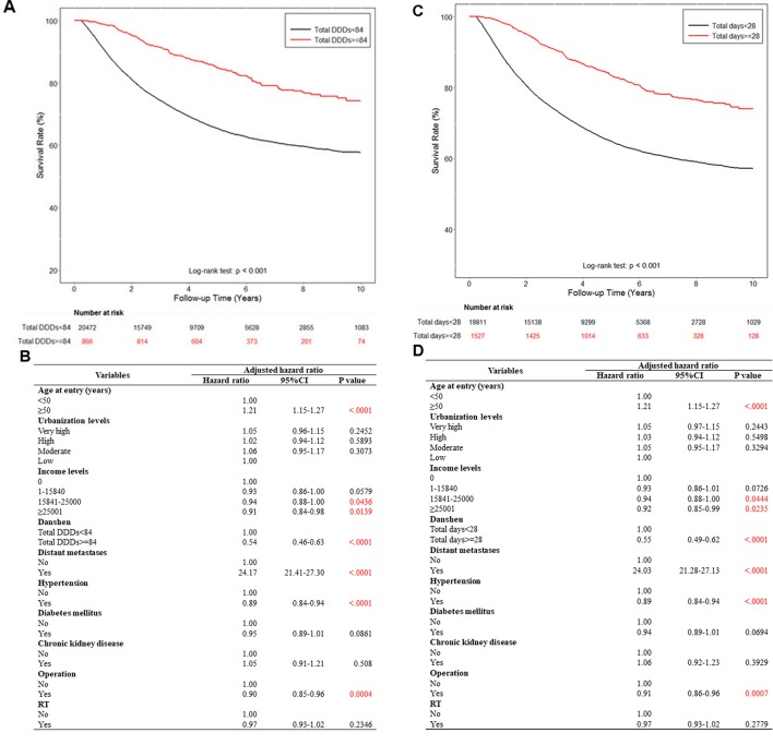
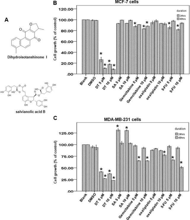
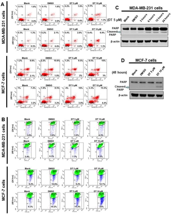
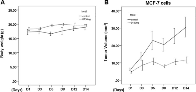
Danshen (salvia miltiorrhiza Bunge) is widely used in traditional Chinese medicine. However, it is definite clinical effort and mechanism on breast cancer is unclear. In our study, we used the real-world database to investigate in vivo protective effort of danshen in the breast cancer patients through using population-based data from the Taiwan National Health Insurance Research Database (NHIRD). In vitro, human breast cancer cells (MCF-7 cells and MDA-MB-231 cells) were used to investigate the effect and the underlying mechanism through XTT assay, flow cytometry, glutathione peroxidase (GPX) activity assay, GSH (reduced glutathione)/GSSG (oxidized glutathione), malondialdehyde (MDA), and western blot analysis. The in vivo effect was investigated through a xenograft nude mouse model. We found that dihydroisotanshinone I (DT), a pure compound present in danshen, can inhibit the growth of breast carcinoma cells, including MCF-7 cells and MDA-MB-231 cells. Moreover, DT induced apoptosis and ferroptosis in these breast cancer cells. DT also repressed the protein expression of GPX4 (Glutathione peroxidase 4). For in vivo study, DT treatment also significantly inhibited the final tumor volume without adverse effects in a xenograft nude mouse model. In conclusion, danshen has protective efforts in breast cancer patients, which could be attributed to DT through inducing apoptosis and ferroptosis of breast cancer cells.

Keywords: GPX4; National Health Insurance Research Database; breast carcinoma; danshen; dihydroisotanshinone I; ferroptosis.


Figures

Figure 1
Figure 1
Patient of breast cancer disposition.
Figure 2
Figure 2
The effect of danshen on the survival rate of breast cancer patients in Taiwan. A total of 79,335 breast cancer patients were included in the study cohort. These patients accrued follow-up time for 10 years. Crude overall Kaplan-Meier survival curves for the breast cancer patients was investigated. (A) The patients were categorized into 2 groups: never used danshen, had used danshen more than 84 grams after breast cancer diagnosed, and those with less than 84 grams danshen used in records. (log-rank: p < 0.001). (C) The patients were categorized into two groups: had used danshen more than 28 days after breast cancer diagnosed and those with less than 28 days with treatment of danshen in records (log-rank: p < 0.001). (B, D) Demographic characteristics of breast cancer patients by medications.
Figure 3
Figure 3
Dihydroisotanshinone I (DT) block the proliferation of breast cancer cell lines. (A) The structure of DT and Salvianolic acid B (SA). (B, C) MCF-7 cells or MDA-MB-231 cells were measured by XTT assay after indicated hours of culturing in the presence of indicated compounds. All the results are representative of at least three independent experiments. (Error bars = mean ± S.E.M. Asterisks (*) mark samples significantly different from DMSO group with p < 0.05).
Figure 4
Figure 4
Dihydroisotanshinone I (DT) induces apoptosis in breast cancer cells. MCF-7 cells or MDA-MB-231 cells were treated without or with indicated compounds for 24–48h. Cell apoptosis was detected by flow cytometry with annexin-V-FITC/PI dual staining or mitoscreen JC-1 staining. (A) For annexin-V-FITC/PI dual staining, the representative histograms of flow cytometric analysis using double staining with annexin-V-FITC (FITC-A) and PI (PI-A). (B) For mitoscreen JC-1 staining, dot Plots revealing depolarization of mitochondria in treated indicated breast cancer cells. The percentage of events in the upper gate (P2) and lower gate (P3) represent population of treated indicated breast cancer cells having normal and depolarized mitochondria respectively. (C, D) Total cell extracts of MDA-MB-231 cells (C) or MCF-7 cells (D) were harvested from cells treated with DMSO or indicated concentrations of DT for indicated hours. The protein was immunoblotted with polyclonal antibodies specific for PARP. β-actin was used as an internal loading control.
Figure 5
Figure 5
DT induced ferroptosis in breast cancer cells in vitro. (A, F) For MDA assay, MCF-7 cells (A) or MDA-MB-231 cells (F) were treated with DMSO or indicated drugs for 24 hours. Total cell extracts of indicate breast cancer cells was collected and analyzed by MDA assay kit. (B) For GPX activity, MCF-7 cells were treated with DMSO or indicated drugs for 24 hours. Total cell extract was collected and analyzed by GPX activity assay kit. (C, G) Total cell extracts of MCF-7 cells (C) or MDA-MB-231 cells (G) were harvested from cells treated with DMSO or indicated concentrations of DT for 24 hours. The protein was immunoblotted with polyclonal antibodies specific for GPX4. β-actin was used as an internal loading control. (D, E, H) For GSH and GSSG level, indicated breast cancer cells were treated with DMSO or indicated drugs for 24 hours. Total cell extract was collected and analyzed by GSH and GSSG assay kit. (Error bars=mean±S.E.M. Asterisks (*) mark samples significantly different from DMSO group with p < 0.05).
Figure 6
Figure 6
The in vivo effect of dihydroisotanshinone I (DT) on xenografted animal model. (A) Average mice weights with every 2-day injection of vehicle/DT over a time course of 2 weeks. (B) Average tumor volume of mice injected with either vehicle (DMSO) or DT (30 mg/kg, n = 5 per group). (Error bars = mean ± S.E.M.).

References

    1. Baysal M., Ilgin S., Kilic G., Kilic V., Ucarcan S., Atli O. (2017). Reproductive toxicity after levetiracetam administration in male rats: Evidence for role of hormonal status and oxidative stress. PLoS One 12, e0175990. 10.1371/journal.pone.0175990 - DOI - PMC - PubMed
    1. Canli O., Alankus Y. B., Grootjans S., Vegi N., Hultner L., Hoppe P. S., et al. (2016). Glutathione peroxidase 4 prevents necroptosis in mouse erythroid precursors. Blood 127, 139–148. 10.1182/blood-2015-06-654194 - DOI - PMC - PubMed
    1. Cardoso F., Costa A., Senkus E., Aapro M., Andre F., Barrios C. H., et al. (2017). 3rd ESO-ESMO International Consensus Guidelines for Advanced Breast Cancer (ABC 3). Ann. Oncol. 28, 16–33. 10.1093/annonc/mdx036 - DOI - PMC - PubMed
    1. De Mello Ramirez Medina J., De Araujo Trugilho I., Mendes G. N. B., Silva J. G., Da Silva Paiva M. A., De Aguiar S. S., et al. (2019). Advanced Clinical Stage at Diagnosis of Breast Cancer Is Associated with Poorer Health-Related Quality of Life: A Cross-Sectional Study. Eur. J. Breast Health 15, 26–31. 10.5152/ejbh.2018.4297 - DOI - PMC - PubMed
    1. Fu P., Du F., Chen W., Yao M., Lv K., Liu Y. (2014). Tanshinone IIA blocks epithelial-mesenchymal transition through HIF-1alpha downregulation, reversing hypoxia-induced chemotherapy resistance in breast cancer cell lines. Oncol. Rep. 31, 2561–2568. 10.3892/or.2014.3140 - DOI - PubMed