

Pancreatic acinar cell carcinoma--literature review and case report of a 56-year-old man presenting with abdominal pain
- PMID: 31737144
- PMCID: PMC6849422
- DOI: 10.1016/j.radcr.2019.10.009
Pancreatic acinar cell carcinoma--literature review and case report of a 56-year-old man presenting with abdominal pain
Erratum in
-
Corrigendum to "Pancreatic acinar cell carcinoma-literature review and case report of a 56-year-old man presenting with abdominal pain" [Radiol Case Rep 15 (2020) 39-43].Radiol Case Rep. 2019 Dec 12;15(3):306-307. doi: 10.1016/j.radcr.2019.11.005. eCollection 2020 Mar. Radiol Case Rep. 2019. PMID: 32071654 Free PMC article.
Abstract
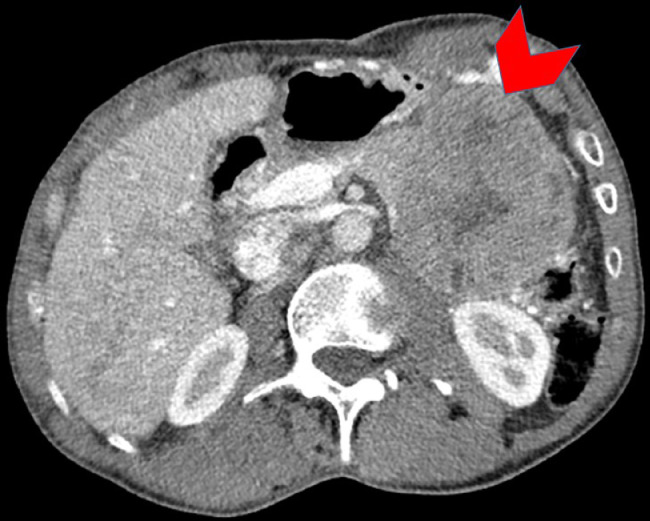
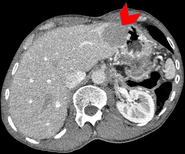
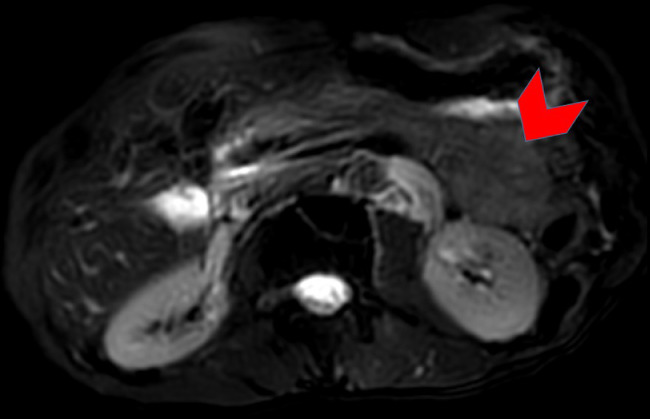
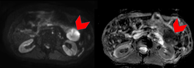
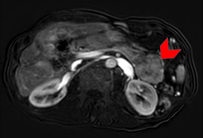
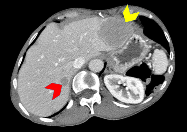
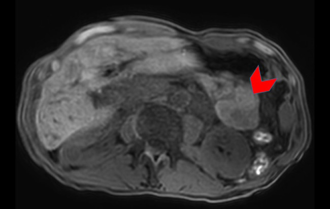
We present a case of acinar cell carcinoma of the pancreas (ACC) with metastasis to the liver in a patient who presented with complaints abdominal pain. The presentation, diagnosis, and management of a 56-year-old man with ACC are discussed here. Imaging with computerized tomography (CT) in particular is crucial in the diagnosis, which can identify the primary lesion as well as metastases. ACC should be considered in the differential as a source of abdominal, epigastric, or back pain with imaging that is suggestive of the diagnosis as prompt recognition and initiation of treatment is paramount in the overall prognosis.
Keywords: Computed tomography; Diagnostic radiology; Magnetic resonance imaging; Oncologic radiology; Pancreatic acinar cell carcinoma.
Published by Elsevier Inc. on behalf of University of Washington.
Figures
References
-
- Solcia E., Capella C., Kloppel G. Tumors of the exocrine pancreas. In: Rosai J., Sorbin L., editors. Atlas of tumor pathology. Armed Forces Institute of Pathology; Washington, DC: 1997. pp. 31–144.
-
- Butturini G, Pisano M, D’Onofrio M, Auriemma A, Bassi C. Aggressive approach to acinar cell carcinoma of the pancreas: a single institution experience and a literature review. Langenbecks Arch Surg. 2011;396(3):363–369. - PubMed
-
- Tatli S., Mortele K. CT and MRI features of pure acinar cell carcinoma of the pancreas in adults. AJR. 2005;184:511–519. - PubMed
Publication types
LinkOut - more resources
Full Text Sources
