. 2019 Nov 12;8(6):3.
doi: 10.1167/tvst.8.6.3. eCollection 2019 Nov.

Presymptomatic Retinal Sensitivity Changes in Intermediate Age-Related Macular Degeneration Associated With New Retinal Fluid

Affiliations

Presymptomatic Retinal Sensitivity Changes in Intermediate Age-Related Macular Degeneration Associated With New Retinal Fluid

Antony J Wightman et al. Transl Vis Sci Technol. .

Abstract

Purpose: To determine whether change in retinal sensitivity in areas with subretinal or intraretinal fluid secondary to age-related macular degeneration (AMD) precedes visual symptoms. If confirmed, retinal sensitivity testing could be used for home monitoring in AMD.

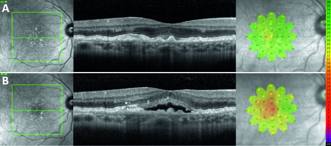
Methods: Individuals with intermediate AMD enrolled in a longitudinal study were seen every 6 months and underwent best-corrected visual acuity testing (BCVA), spectral domain-optical coherence tomography (SD-OCT), and microperimetry. Asymptomatic individuals who developed incidental, reading center-determined retinal fluid detected on SD-OCT were identified. The point-wise sensitivity (PWS) at the time of fluid detection was compared with 6 and 12 months prior.

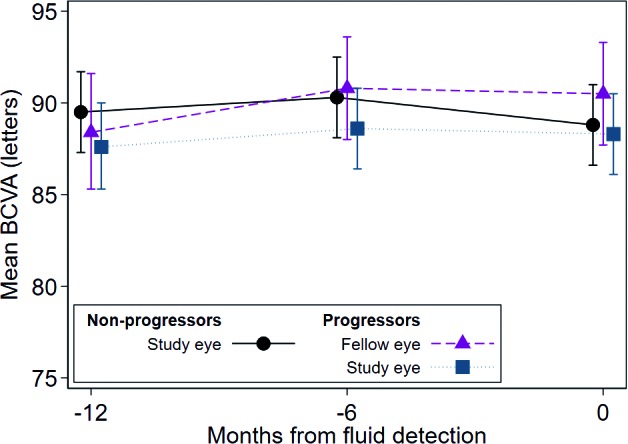
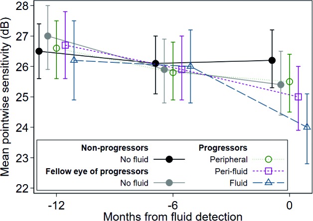
Results: Fourteen of 161 individuals developed fluid without symptoms. PWS over fluid areas at detection was reduced compared with 6 (difference -2.04 dB, P < 0.001) and 12 months (-2.27 dB, P < 0.001) prior. PWS over fluid areas was reduced compared with perifluid areas (difference -1.02 dB, P = 0.03), peripheral areas (-1.51 dB, P < 0.001), nonprogressed fellow eyes (-1.49 dB, P = 0.006), and nonprogressed age-matched intermediate AMD eyes (-2.29 dB, P = 0.001). No difference in BCVA was observed in eyes developing fluid compared to eyes that do not develop fluid (P = 0.76).

Conclusions: Retinal areas with fluid on SD-OCT had a corresponding reduction in retinal sensitivity at the time of fluid detection compared with 6 and 12 months prior, in asymptomatic intermediate AMD without change in BCVA.

Translational relevance: Development of self-monitoring tools to detect changes in retinal sensitivity may be helpful for early detection of retinal fluid suggestive of progression to neovascular AMD before acuity is affected.

Keywords: SD-OCT; age-related macular degeneration; microperimetry; retinal fluid; retinal sensitivity.


Figures

Figure 1
Figure 1
(A) Retinal thickness heat map generated by the Eye Explorer software (Heidelberg Engineering) demonstrating an area of elevation (red) secondary to retinal fluid at the inferior nasal retina of a left eye. False-color scale bar shows retinal thickness (μm). (B) NIR scout image with overlaid retinal sensitivity results from the MAIA. The AMD 6° grid is colored to denote varying threshold measurement values (dB) showing the area of low sensitivity is associated with retinal fluid. (C) Relationship between the fluid location (yellow boundary) and the location of the sensitivity testing (orange circles). Points falling within or on the area demarcated by the yellow boundary were classified as co-located with fluid (yellow-filled test points), those immediately surrounding were classified as perifluid (white-filled test points) and those further removed were classified as peripheral to fluid.
Figure 2
Figure 2
Longitudinal changes in mean BCVA (letters) of nonprogressor and progressor groups for three visits shows no change in mean BCVA over time for any group. Estimated using mixed-effects piecewise linear regression model with random intercepts, accounting for correlation within eye and participant and adjusted for age and smoking status. Error bars indicate 95%CI. See Table 2 for statistical analysis. Note that individual data points have a slight spread at the three longitudinal time points so that each data point is visible. All data points were obtained at the time of fluid detection and at 6 and 12 months prior.
Figure 3
Figure 3
Mean pointwise sensitivity estimated using a mixed-effects piecewise linear regression model with random intercepts, accounting for correlation within eye, participant and stimulus location, and adjusted for age and smoking status. Note the greatest decrease in sensitivity demonstrated by areas overlying fluid at time of fluid detection. Error bars indicate 95%CI. See Table 3 for statistical analysis. Note that individual data points have a slight spread at the three longitudinal time points so that each data point is visible. All data points were obtained at the time of fluid detection and at 6 and 12 months prior.
Figure 4
Figure 4
Representative finding from a 70-year-old female study participant who developed SRF inferonasally to the fovea in their left eye. (A) NIR scout image, SD-OCT B-scan, and MAIA findings 6 months prior to development of SRF. (B) NIR, SD-OCT, and MAIA at the time of SRF detection. Microperimetry scale of decibel values and corresponding colors is shown on the right.
Figure 5
Figure 5
Representative finding from a 64-year-old female study participant who developed SRF centrally in their right eye. (A) NIR scout image, SD-OCT B-scan, and MAIA findings 6 months prior to development of SRF. (B) NIR, SD-OCT, and MAIA at the time of SRF detection. Microperimetry scale of decibel values and corresponding colors is shown on the right.

References

    1. Bloch SB, Larsen M, Munch IC. Incidence of legal blindness from age-related macular degeneration in Denmark: year 2000 to 2010. Am J Ophthalmol. 2012;153:209–213. e202. - PubMed
    1. Mitchell P, Bressler N, Doan QV, et al. Estimated cases of blindness and visual impairment from neovascular age-related macular degeneration avoided in Australia by ranibizumab treatment. PLoS One. 2014;9:e101072. - PMC - PubMed
    1. Fong DS, Custis P, Howes J, Hsu J-W. Intravitreal bevacizumab and ranibizumab for age-related macular degeneration: a multicenter, retrospective study. Ophthalmology. 2010;117:298–302. - PubMed
    1. Olsen TW, Feng X, Kasper TJ, Rath PP, Steuer ER. Fluorescein angiographic lesion type frequency in neovascular age-related macular degeneration. Ophthalmology. 2004;111:250–255. - PubMed
    1. Finger RP, Wickremasinghe SS, Baird PN, Guymer RH. Predictors of anti-VEGF treatment response in neovascular age-related macular degeneration. Surv Ophthalmol. 2014;59:1–18. - PubMed