Review
. 2019 Oct;9(Suppl 2):S388-S415.
doi: 10.21037/cdt.2019.02.01.

Hypertrophic cardiomyopathy: genetics and clinical perspectives

Affiliations
Review

Hypertrophic cardiomyopathy: genetics and clinical perspectives

Cordula Maria Wolf. Cardiovasc Diagn Ther. 2019 Oct.

Abstract

Hypertrophic cardiomyopathy (HCM) is the most common inherited heart disease and defined by unexplained isolated progressive myocardial hypertrophy, systolic and diastolic ventricular dysfunction, arrhythmias, sudden cardiac death and histopathologic changes, such as myocyte disarray and myocardial fibrosis. Mutations in genes encoding for proteins of the contractile apparatus of the cardiomyocyte, such as β-myosin heavy chain and myosin binding protein C, have been identified as cause of the disease. Disease is caused by altered biophysical properties of the cardiomyocyte, disturbed calcium handling, and abnormal cellular metabolism. Mutations in sarcomere genes can also activate other signaling pathways via transcriptional activation and can influence non-cardiac cells, such as fibroblasts. Additional environmental, genetic and epigenetic factors result in heterogeneous disease expression. The clinical course of the disease varies greatly with some patients presenting during childhood while others remain asymptomatic until late in life. Patients can present with either heart failure symptoms or the first symptom can be sudden death due to malignant ventricular arrhythmias. The morphological and pathological heterogeneity results in prognosis uncertainty and makes patient management challenging. Current standard therapeutic measures include the prevention of sudden death by prohibition of competitive sport participation and the implantation of cardioverter-defibrillators if indicated, as well as symptomatic heart failure therapies or cardiac transplantation. There exists no causal therapy for this monogenic autosomal-dominant inherited disorder, so that the focus of current management is on early identification of asymptomatic patients at risk through molecular diagnostic and clinical cascade screening of family members, optimal sudden death risk stratification, and timely initiation of preventative therapies to avoid disease progression to the irreversible adverse myocardial remodeling stage. Genetic diagnosis allowing identification of asymptomatic affected patients prior to clinical disease onset, new imaging technologies, and the establishment of international guidelines have optimized treatment and sudden death risk stratification lowering mortality dramatically within the last decade. However, a thorough understanding of underlying disease pathogenesis, regular clinical follow-up, family counseling, and preventative treatment is required to minimize morbidity and mortality of affected patients. This review summarizes current knowledge about molecular genetics and pathogenesis of HCM secondary to mutations in the sarcomere and provides an overview about current evidence and guidelines in clinical patient management. The overview will focus on clinical staging based on disease mechanism allowing timely initiation of preventative measures. An outlook about so far experimental treatments and potential for future therapies will be provided.

Keywords: Hypertrophic cardiomyopathy (HCM); clinical management; molecular genetics; pathogenesis; sudden cardiac death.


Conflict of interest statement

Conflicts of Interest: The author has no conflicts of interest to declare.

Figures

Figure 1
Figure 1
Differential diagnosis of unexplained left ventricular hypertrophy. Left ventricular hypertrophy (LVH) can present isolated or be associated with an underlying multisystem disorder. The diagram presents an overview of distinct etiologies causing LVH. The proposed clinical work-up applies for adult patients and is incorporated as class I and class IIA indications according to ESC (32) and AHA (18) guidelines. Establishing the correct diagnosis in a patient presenting with LVH is crucial to offer specific treatment and counseling. Genetic and clinical family screening is important if a diagnosis of hypertrophic cardiomyopathy (HCM) is made to identify asymptomatic disease carriers. ECG, electrocardiogram; TTE, transthoracic echocardiography; MELAS, mitochondrial encephalopathy, lactic acidosis, and stroke-like episodes; MERFF, myoclonic epilepsy with ragged red fiber; CFC, cardiofaciocutaneous syndrome.
Figure 2
Figure 2
Pathogenesis of hypertrophic cardiomyopathy. The primary defect is the mutation in a gene encoding for a sarcomere protein. This mutation alters protein expression, morphology and function and influences sarcomere assembly, force generation, intracellular calcium homeostasis, and ATPase activity. Those altered biophysical and intracellular properties cause cardiomyocyte hypertrophy, diastolic and systolic dysfunction, rhythm disturbances and histopathologic changes over time. The underlying gene defect initiates secondary molecular changes, such as pro-hypertrophic and pro-fibrotic signaling (e.g., TGF-β, Periostin, MEF2, MAPK, RAS, NFAT), gene expression, post-translational modifications, mitochondrial dysfunction and mitotic factors. Those changes lead to the pathognomic histopathological changes, such as myocyte disarray and myocardial fibrosis. Arrhythmias can occur as a sequela of both disturbed intracellular calcium handling, cardiomyocyte hypertrophy and myocardial histopathological changes. Other pathogenic genetic variants (modifiers), genomic/epigenetic factors (e.g., miRNA, long non-coding RNA, histon modification), proteomics (e.g., post-translational modifications, cell-cell-communication) and environmental factors contributing to expression of the phenotype and result in large clinical variability.
Figure 3
Figure 3
Pathomechanisms and associated clinical findings of arrhythmias in hypertrophic cardiomyopathy. Abnormal automaticity, triggered activity, and reentry promote ventricular arrhythmia which can ultimately cause sudden cardiac death. In hypertrophic cardiomyopathy, cardiomyocyte stretch is believed to increase automaticity by influencing self-depolarizing channels, such as the funny channel If and the T-type calcium channel ICaT. Altered calcium homeostasis and altered biophysical properties can initiate early and late afterdepolarizations, presenting as premature ventricular contractions on Holter monitoring, which in turn can initiate non-sustained and sustained ventricular arrhythmias. Myocyte disarray, focal and interstitial fibrosis provide the anatomical substrate that predisposes to conduction block and promote macro- and microreentry arrhythmias. Multiple transient pathophysiological factors, such as altered hemodynamics, autonomic responses, or myocardial ischemia modulate the arrhythmogenic potential favoring electrical triggers.
Figure 4
Figure 4
Clinical disease stages and stage-specific clinical management of hypertrophic cardiomyopathy. Hypertrophic cardiomyopathy (HCM) is a congenital heart defect, but disease progression occurs over life and medical management needs to be adopted to disease stage. Molecular testing can diagnose the underlying genetic defect before the disease becomes overt in a patient (Genotype+/Phenotype−, stage 0, green boxes). Regular cardiac evaluation and appropriate family counseling needs to be performed during this stage. The frequency of cardiac screening should be based on family history, current guidelines, and healthcare provider recommendation. Stage I presents with myocardial hypertrophy in asymptomatic or mildly symptomatic patients with subtle clinical findings (yellow boxes). Treatment of left ventricular outflow tract obstruction and thorough risk stratification for sudden arrhythmic death is required during this stage. Regular cardiac evaluation should be performed to timely identify progression to the “adverse remodeling” stage (stage II, orange boxes). More aggressive treatment is typically required during this stage. Specific emphasis should be made on new therapeutic options preventing disease progression to avoid progression to end-stage disease (stage III, red boxes). Stage III presents usually an irreversible disease process in highly symptomatic patients. There is high morbidity and mortality during this stage.
Figure 5
Figure 5
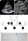
Typical clinical findings of patients with hypertrophic cardiomyopathy. Left ventricular (LV) hypertrophy (white stars) and left atrial (LA) enlargement can be best detected by transthoracic echocardiography (panel A). Focal myocardial fibrosis is diagnosed by positive late gadolinium enhancement (white arrows) on cardiac magnetic resonance tomography (panel B). Panel C: high incidence of sudden cardiac death or appropriate discharge of the implantable cardioverter defibrillator (ICD, black arrow on intracardiac electrogram panel D) in a family carrying a mutation in the β-myosin heavy chain (MYH7) and the cardiac troponin T (TNNT2) gene (black filled symbols, panel C).

References

    1. Semsarian C, Ingles J, Maron MS, et al. New perspectives on the prevalence of hypertrophic cardiomyopathy. J Am Coll Cardiol 2015;65:1249-54. 10.1016/j.jacc.2015.01.019 - DOI - PubMed
    1. Maron BJ, Gardin JM, Flack JM, et al. Prevalence of hypertrophic cardiomyopathy in a general population of young adults. Echocardiographic analysis of 4111 subjects in the CARDIA Study. Coronary Artery Risk Development in (Young) Adults. Circulation 1995;92:785-9. 10.1161/01.CIR.92.4.785 - DOI - PubMed
    1. Braunwald E, Lambrew CT, Rockoff SD, et al. Idiopathic Hypertrophic Subaortic Stenosis. I. A Description of the Disease Based Upon an Analysis of 64 Patients. Circulation 1964;30:SUPPL 4:4-119. - PubMed
    1. Elliott P, Andersson B, Arbustini E, et al. Classification of the cardiomyopathies: a position statement from the European Society Of Cardiology Working Group on Myocardial and Pericardial Diseases. Eur Heart J 2008;29:270-6. 10.1093/eurheartj/ehm342 - DOI - PubMed
    1. Maron BJ, Towbin JA, Thiene G, et al. Contemporary definitions and classification of the cardiomyopathies: an American Heart Association Scientific Statement from the Council on Clinical Cardiology, Heart Failure and Transplantation Committee; Quality of Care and Outcomes Research and Functional Genomics and Translational Biology Interdisciplinary Working Groups; and Council on Epidemiology and Prevention. Circulation 2006;113:1807-16. 10.1161/CIRCULATIONAHA.106.174287 - DOI - PubMed