Case Reports
. 2019 Aug 8;6(8):e00154.
doi: 10.14309/crj.0000000000000154. eCollection 2019 Aug.

Gastric Ulcer and Perforation due to Mucormycosis in an Immunocompetent Patient

Affiliations
Case Reports

Gastric Ulcer and Perforation due to Mucormycosis in an Immunocompetent Patient

Gurbir Sehmbey et al. ACG Case Rep J. .

Abstract

Mucormycosis is a rare and life-threatening fungal infection that is associated with high mortality in immunocompromised individuals. Although it most commonly affects lungs and paranasal sinuses, cases of invasive mucormycosis of the gastrointestinal tract have also been reported. Gastrointestinal mucormycosis (GIM) is most commonly found in the stomach, colon, and ileum. Etiologies of GIM include ingestion of spores and penetrating abdominal trauma, causing mucocutaneous disruption. We present a case of an immunocompetent man who presented to our hospital after a gunshot wound to the abdomen. His hospital course was complicated with the development of invasive GIM in the form of a large gastric ulcer, which caused gastrointestinal bleeding and eventually perforation.


Figures

Figure 1.
Figure 1.
(A and B) Large gastric ulcer with exudate within the body and fundus of the stomach. An overlying feeding tube can also be seen.
Figure 2.
Figure 2.
(A) Hematoxylin & eosin (H&E) stain of gastric ulcer biopsy at low magnification showing necrotic exudate containing fungal aseptate hyphae, morphologically consistent with mucormycosis. (B) H&E stain of gastric ulcer biopsy at medium magnification showing fungal aseptate hyphae, consistent with invasive mucormycosis.
Figure 3.
Figure 3.
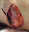
Necrotic lesion at the open left flank ballistic wound.

References

    1. Lelievre L, Garcia-Hermoso D, Abdoul H, et al. Posttraumatic mucormycosis: A nationwide study in France and review of the literature. Medicine (Baltimore). 2014;93(24):395–404. - PMC - PubMed
    1. Bitar D, Van Cauteren D, Lanternier F, et al. Increasing incidence of zygomycosis (mucormycosis), France, 1997e2006. Emerg Infect Dis. 2009;15:1395e401. - PMC - PubMed
    1. Kontoyiannis DP, Lewis RE, Invasive zygomycosis: Update on pathogenesis, clinical manifestations, and management. Infect Dis Clin North Am. 2006;20(3):581–607. - PubMed
    1. Sugar AM. Mucormycosis. Clin Infect Dis. 1992;14:5126–9. - PubMed
    1. Spellberg B. Gastrointestinal mucormycosis: An evolving disease, Gastroenterol Hepatol. 2012;8(2):140–2. - PMC - PubMed

Publication types