

The retinal choroid as an oculovascular biomarker for Alzheimer's dementia: A histopathological study in severe disease
- PMID: 31737776
- PMCID: PMC6849152
- DOI: 10.1016/j.dadm.2019.08.005
The retinal choroid as an oculovascular biomarker for Alzheimer's dementia: A histopathological study in severe disease
Abstract
Introduction: Previous in vivo optical coherence tomography studies have proposed the retinal choroid as a potential oculovascular biomarker for Alzheimer's disease (AD). However, the clinical use of the choroid as a purported surrogate marker remains poorly understood. We pursued a histopathological approach to assess choroidal thickness and vascular morphology in severe disease.
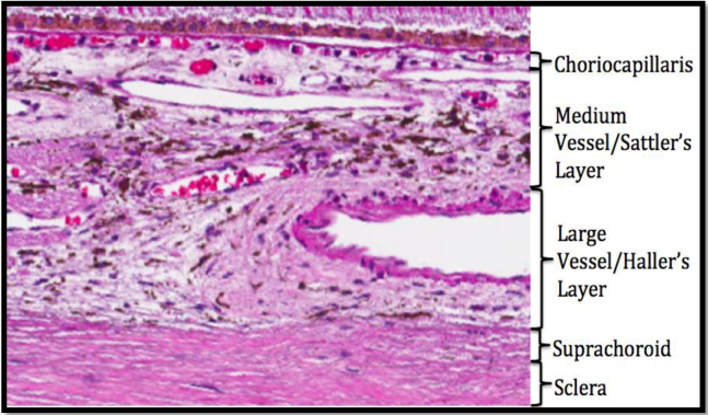
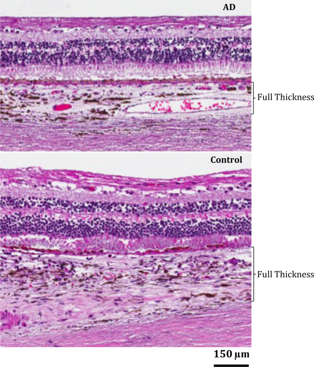
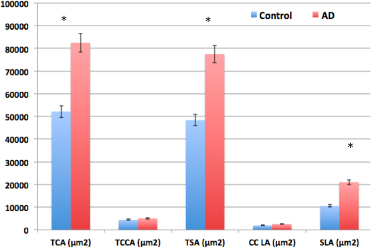
Methods: Human postmortem tissues from 8 patients with AD (mean age: 80.1 ± 12.7 years) and from 11 age-matched controls (mean age: 78.4 ± 16.57 years) were analyzed. Thickness, area, and vascularity of the retinal choroid and its sublayers were measured from the nasal and temporal quadrants of the superior retina.
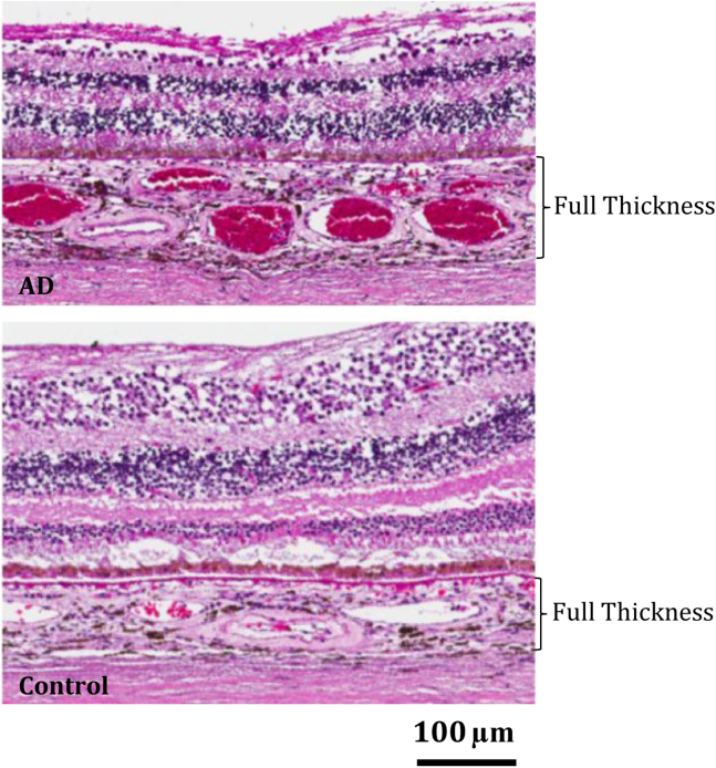
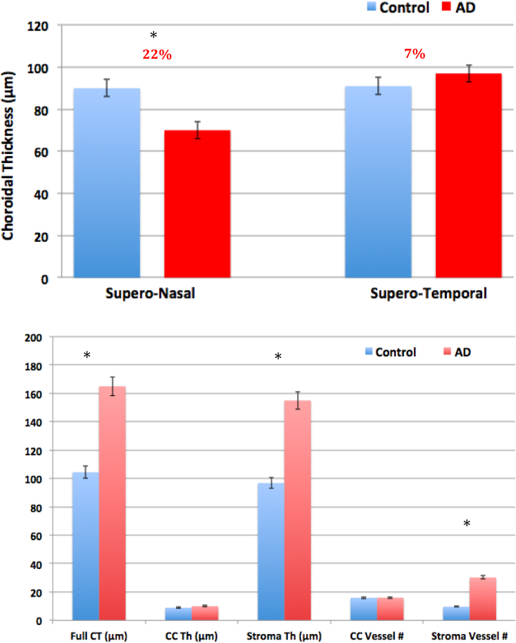
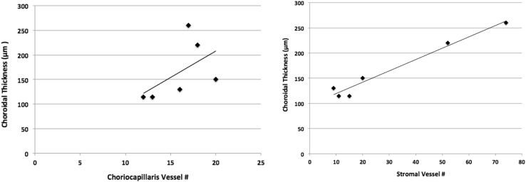
Results: Nasally, the choroid was thinner in the patients with AD than in the controls (22% thickness reduction; P < .001), but to our surprise, the choroid was thicker in the patients with AD than in the controls (~60% increase; P < .03) within the macula, temporally. The choroidal area was also significantly greater in the patients with AD than in the controls (~60% increase; P < .03). Choroidal thickening in AD was strongly correlated with the stromal vessel number (R2 = 0.96, P < .001).
Discussion: We found significant differences in the retinal choroid by layer and by region, nasally and temporally with respect to the optic nerve. Intriguingly, the choroid was markedly thicker in the central macular region and was strongly associated with vessel number in the stromal vascular layer. These quantified histological findings in severe disease expand our understanding of vascular pathology in AD and suggest vascularity as a potential biomarker supplementary to thickness when evaluating the retinal choroid in AD.
Keywords: Alzheimer's disease; Biomarker; Choroid; Histopathology; Morphometric analysis; Retina; Vascular biomarkers.
© 2019 The Authors.
Figures
References
-
- 2019 Alzheimer's disease facts and figures. Alzheimers Dement. 2019;15:321–387.
-
- Pillai J.A., Cummings J.L. Clinical trials in predementia stages of Alzheimer disease. Med Clin North Am. 2013;97:439–457. - PubMed
-
- Katz B., Rimmer S. Ophthalmologic manifestations of Alzheimer's disease. Surv Ophthalmol. 1989;34:31–43. - PubMed
-
- Uhlmann R.F., Larson E.B., Koepsell T.D., Rees T.S., Duckert L.G. Visual impairment and cognitive dysfunction in Alzheimer's disease. J Gen Intern Med. 1991;6:126–132. - PubMed
-
- Salamone G., Di lorenzo C., Mosti S., Lupo F., Cravello L., Palmer K. Color discrimination performance in patients with Alzheimer's disease. Dement Geriatr Cogn Disord. 2009;27:501–507. - PubMed