Review
. 2020 Apr;95(2):365-392.
doi: 10.1111/brv.12569. Epub 2019 Nov 18.

Post-ejaculatory modifications to sperm (PEMS)

Affiliations
Review

Post-ejaculatory modifications to sperm (PEMS)

Scott Pitnick et al. Biol Rev Camb Philos Soc. 2020 Apr.

Abstract

Mammalian sperm must spend a minimum period of time within a female reproductive tract to achieve the capacity to fertilize oocytes. This phenomenon, termed sperm 'capacitation', was discovered nearly seven decades ago and opened a window into the complexities of sperm-female interaction. Capacitation is most commonly used to refer to a specific combination of processes that are believed to be widespread in mammals and includes modifications to the sperm plasma membrane, elevation of intracellular cyclic AMP levels, induction of protein tyrosine phosphorylation, increased intracellular Ca2+ levels, hyperactivation of motility, and, eventually, the acrosome reaction. Capacitation is only one example of post-ejaculatory modifications to sperm (PEMS) that are widespread throughout the animal kingdom. Although PEMS are less well studied in non-mammalian taxa, they likely represent the rule rather than the exception in species with internal fertilization. These PEMS are diverse in form and collectively represent the outcome of selection fashioning complex maturational trajectories of sperm that include multiple, sequential phenotypes that are specialized for stage-specific functionality within the female. In many cases, PEMS are critical for sperm to migrate successfully through the female reproductive tract, survive a protracted period of storage, reach the site of fertilization and/or achieve the capacity to fertilize eggs. We predict that PEMS will exhibit widespread phenotypic plasticity mediated by sperm-female interactions. The successful execution of PEMS thus has important implications for variation in fitness and the operation of post-copulatory sexual selection. Furthermore, it may provide a widespread mechanism of reproductive isolation and the maintenance of species boundaries. Despite their possible ubiquity and importance, the investigation of PEMS has been largely descriptive, lacking any phylogenetic consideration with regard to divergence, and there have been no theoretical or empirical investigations of their evolutionary significance. Here, we (i) clarify PEMS-related nomenclature; (ii) address the evolutionary origin, maintenance and divergence in PEMS in the context of the protracted life history of sperm and the complex, selective environment of the female reproductive tract; (iii) describe taxonomically widespread types of PEMS: sperm activation, chemotaxis and the dissociation of sperm conjugates; (iv) review the occurence of PEMS throughout the animal kingdom; (v) consider alternative hypotheses for the adaptive value of PEMS; (vi) speculate on the evolutionary implications of PEMS for genomic architecture, sexual selection, and reproductive isolation; and (vii) suggest fruitful directions for future functional and evolutionary analyses of PEMS.

Keywords: capacitation; female reproductive tract; fertility; hyperactivation; morphogenesis; motility; post-copulatory sexual selection; seminal proteins; sperm competition; spermatozoa.


Figures

Fig. 1.
Fig. 1.
Sperm conjugates of the whirligig beetle, Dineutus sp.: (A) conjugates from the male ejaculatory duct under differential interference contrast microscopy; (B) conjugates stained with 4′,6-diamidino-2-phenylindole (DAPI) to show the organization of sperm heads along the spermatostyle; (C) conjugate from the female spermatheca in the process of sperm dissociation; (D) bundle of spermless spermatostyles from the spermatheca of a wild-caught female. Photomicrographs by S. Pitnick.
Fig. 2.
Fig. 2.
Tranmission electron micrographs showing post-ejaculatory modifications to sperm (PEMS) in the form of digitate processes (examples indicated by arrows) formed from the sperm periacrosomal plasmalemma in order to strengthen contact with (A, B) the female spermathecal cell membrane in the polychaete worm, Spirorbis spirorbis, and (C, D) the female gill filament in the brooding clam, Mysella tumida. a, acrosome; f, flagellum; m, mitochondrion; n, nucleus; *, specialized contacts between sperm and spermathecal cell membranes with scalariform junctions. Adopted with permission from (A, B) Daly & Golding (1977); (C, D) Ó Foighil (1985b). Scale bars: A, 0.5 μm; C, 2 μm; D, 0.4 μm.
Fig. 3.
Fig. 3.
Scanning electron micrographs of Octopus vulgaris spermatozoon collected from (A) the spermatophore, with an intact outer membrane covering the acrosome, and (B) from the female oviducal gland, following post-ejaculatory modifications to sperm (PEMS) to reveal the corkscrew-shaped acrosome. Inset in A shows magnification of the acrosomal region. Arrows indicate indentations separating the acrosome, nuclear and midpiece regions. Adopted with permission from Tosti et al. (2001). Scale bars: A, 3.0 μm; B, 2.0 μm.
Fig. 4.
Fig. 4.
Schematic diagram of different morphological stages of post-ejaculatory modifications to sperm (PEMS) of the tick, Amblyomma dissimili. mp, motile processes. Adopted with permission from Reger (1963).
Fig. 5.
Fig. 5.
Sperm of the spider Caponina alegre (A–D) before and (E) after post-ejaculatory modifications to sperm (PEMS). (A–D) Reconstruction of a synspermium. The image stack used for the three-dimensional reconstruction is stored in MorphDBase (https://www.morphdbase.de?P_Michalik_20120927-M-3.1). (A) Numerous membrane-bound vesicles are attached to the vesicular area, enclosing the spermatozoa. (B) The main cell components of the four fused spermatozoa are coiled within the vesicular area of the syncytium, but not twisted around each other. (C) The prominent extremely elongated nucleus of one spermatozoon is coiled 2.5 times around the centre of the syncytium into which the axoneme finally opens. (D) Cross-section through a synspermium showing the arrangement of the coiled sperm components in the periphery of the syncytium, leaving the centre only filled with the vesicular area. (E) Schematic drawing of the main components of a post-PEMS, mature spermatozoon. AC, acrosomal complex (acrosomal vacuole and acrosomal filament); AF, acrosomal filament; Ax, axoneme; IF, implantation fossa; peN, post-centriolar elongation of nucleus; prcN, precentriolar region of nucleus. Adopted with permission from Lipke & Michalik (2012).
Fig. 6.
Fig. 6.
(A) Scanning electron micrograph (SEM) of two rolled spermatozoa showing the long extra-acrosomal structure (‘peduncle’) of the collembolan Allacma fusca. Inset, SEM of a single rolled sperm showing the acrosome and peduncle. (B) Schematic reconstruction of a spermatozoon of Orchesella villosa. Sperm components form several spires within the same plasma membrane surrounding material within an ‘extracellular’ cavity. A, acrosome; EAS, extra-acrosomal structure; Ex, extracellular cavity; sp, spermatozoon. Adopted with permission from (A) Fanciulli et al. (2017); (B) Dallai et al. (2004).
Fig. 7.
Fig. 7.
Schematic drawing of a spermatozoon of the jumping bristletail, Machilis distincta, from the female spermatheca but prior to post-ejaculatory modifications to sperm (PEMS). Adopted with permission from Dallai (1972).
Fig. 8.
Fig. 8.
(A) Schematic of a ‘mature’ sperm from the testis (top) and the female spermatheca 2 days after insemination (bottom) in the fungus gnat, Sciara coprophila. Discontinuities in the diagrams indicate that the cell is much longer relative to the width than depicted. (B) Changes undergone by the axial filament complex during storage in the female reproductive tract. (C, D) Transmission electron micrograph of transverse section through the subnuclear portion of a ‘mature’ sperm from (C) the male testis and (D) the female spermatheca. A, acrosome; AF, axial filament complex; B, dense body; MC, mitochondrial crystalloid; MH, mitochondrial homogeneous material; N, nucleus. Adopted with permission from (A, B, D) Phillips (1966a); (C) Dallai, Bernini, & Giusti (1973).
Fig. 9.
Fig. 9.
A model of molecular post-ejaculatory modifications to sperm (PEMS) in Drosophila melanogaster. A network of seminal proteins is required for sex peptide (SP) to bind stably to sperm within the female seminal receptacle. Coloured shapes indicate proteins produced in the male accessory glands. CG1652 and CG1656 require fellow network proteins CG9997 and Antr to be transferred to females. Once deposited in females, Sems and CG17575 are required for SP and CG1656 to localize to the seminal receptacle (SR), the major site of female sperm storage. In the SR, SP and CG1656 bind sperm within 2 h of the start of mating. Also, within the female reproductive tract (FRT), the presence of CG1652 and CG1656 slows the rate at which CG9997 is processed from a 45 kDa form to a 36 kDa form. One additional network protein, Intrepid, is not shown, since its position in the pathway is presently unknown. Loss of any one of these network proteins prevents SP accumulation on sperm in the SR. Following the events shown, the SP C-terminus is cleaved from stored sperm over time. Colours indicate predicted protein functional classes: red/ orange/yellow are serine proteases and protease homologs; pink/purple are cysteine-rich secretory proteins; green are C-type lectins. Adopted with permission from Singh et al. (2018).
Fig. 10.
Fig. 10.
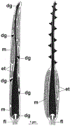
Schematic drawing illustrating post-ejaculatory modifications to sperm (PEMS) of the tunicate, Diplosoma listerianum. Head of a spermatozoon from the male’s sperm duct (left) and from the female’s ovarian fertilization canal (right). et, endoplasmic tubules; fl, flagellum; m, mitochondrion; dg, dense groove. Adopted with permission from Burighel & Martinucci (1994a).
Fig. 11.
Fig. 11.
Schematic illustrating post-ejaculatory modifications to the head of the spermatozoon of the Chinese soft-shelled turtle, Pelodiscus sinensis. Adopted with permission from Zhang et al. (2015).
Fig. 12.
Fig. 12.
Differential interference contrast micrographs of (A, B) many sperm stored within the deep portion of an isthmic crypt, and of (C, D) a single sperm released from storage of a female of the dasyurid marsupial, Sminthopsis crassicaudata, mated about 24–26 h previously. (A, C) The female is preovulatory and all sperm are spear shaped, with the anterior midpiece of the tail lying within lateral folds of the head; (B, D) the female is post-ovulatory and all sperm are T-shaped with the head angulated or perpendicular to the tail (the flagellum in panel D was oscillating and so appears blurred). Adopted with permission from Bedford & Breed (1994).

References

    1. Fuentes-G JA, Housworth EA, Weber A & Martins EP (2016). Phylogenetic ANCOVA: estimating changes in evolutionary rates as well as relationships between traits. The American Naturalist 188, 615–627. - PubMed
    1. Aalberts M, Stout TAE & Stoorvogel W, (2014). Prostasomes: extracellular vesicles from the prostate. Reproduction 147, R1–R14. - PubMed
    1. Adams DC (2014). Quantifying and comparing phylogenetic evolutionary rates for shape and other high-dimensional phenotypic data. Systematic Biology 63, 166–177. - PubMed
    1. Afzelius BA & Dallai R (1987). Conjugated spermatozoa In New Horizons in Sperm Cell Research (ed. Mohri H), pp. 349–355. Japan Science Society, Tokyo.
    1. Ahmed-Braimah YH & McAllister BF (2012). Rapid evolution of assortative fertilization between recently allopatric species of Drosophila. International Journal of Evolutionary Biology 2012, 285468. - PMC - PubMed

Publication types