. 2020 Jan 15;201(2):198-211.
doi: 10.1164/rccm.201902-0451OC.

Grp78 Loss in Epithelial Progenitors Reveals an Age-linked Role for Endoplasmic Reticulum Stress in Pulmonary Fibrosis

Affiliations

Grp78 Loss in Epithelial Progenitors Reveals an Age-linked Role for Endoplasmic Reticulum Stress in Pulmonary Fibrosis

Zea Borok et al. Am J Respir Crit Care Med. .

Abstract

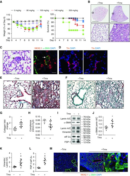
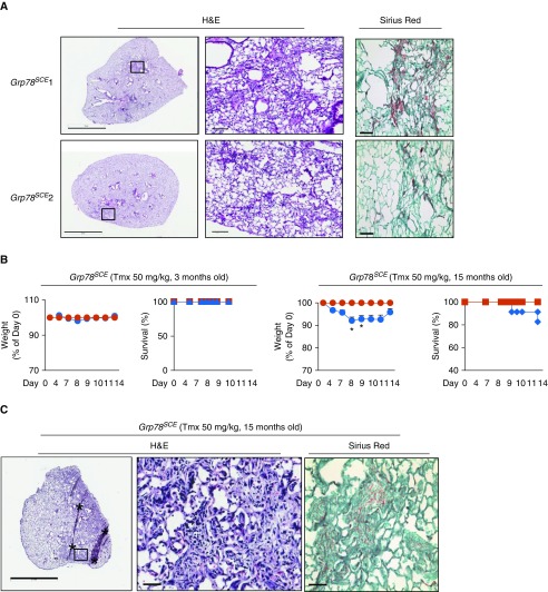
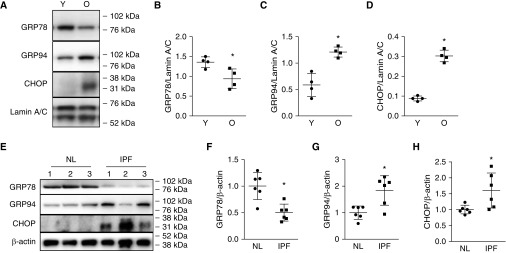
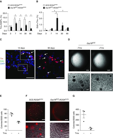
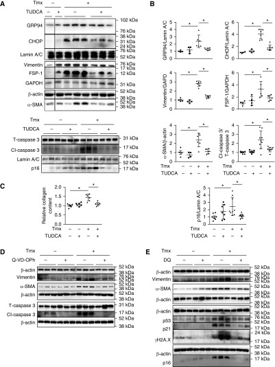
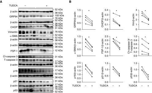
Rationale: Alveolar epithelial cell (AEC) injury and dysregulated repair are implicated in the pathogenesis of pulmonary fibrosis. Endoplasmic reticulum (ER) stress in AEC has been observed in idiopathic pulmonary fibrosis (IPF), a disease of aging.Objectives: To investigate a causal role for ER stress in the pathogenesis of pulmonary fibrosis (PF) and therapeutic potential of ER stress inhibition in PF.Methods: The role of ER stress in AEC dysfunction and fibrosis was studied in mice with tamoxifen (Tmx)-inducible deletion of ER chaperone Grp78, a key regulator of ER homeostasis, in alveolar type II (AT2) cells, progenitors of distal lung epithelium, and in IPF lung slice cultures.Measurements and Main Results:Grp78 deletion caused weight loss, mortality, lung inflammation, and spatially heterogeneous fibrosis characterized by fibroblastic foci, hyperplastic AT2 cells, and increased susceptibility of old and male mice, all features of IPF. Fibrosis was more persistent in more severely injured Grp78 knockout (KO) mice. Grp78 KO AT2 cells showed evidence of ER stress, apoptosis, senescence, impaired progenitor capacity, and activation of TGF-β (transforming growth factor-β)/SMAD signaling. Glucose-regulated protein 78 is reduced in AT2 cells from old mice and patients with IPF, and ER stress inhibitor tauroursodeoxycholic acid ameliorates ER stress and fibrosis in Grp78 KO mouse and IPF lung slice cultures.Conclusions: These results support a causal role for ER stress and resulting epithelial dysfunction in PF and suggest ER stress as a potential mechanism linking aging to IPF. Modulation of ER stress and chaperone function may offer a promising therapeutic approach for pulmonary fibrosis.

Keywords: ER stress; alveolar epithelial cell dysfunction; pulmonary fibrosis.


Figures

Figure 1.
Figure 1.
Grp78 (glucose-regulated protein 78) knockout (KO) induces fibrosis in Grp78SCE mice. (A) Tamoxifen (Tmx) injection (80, 100, 140, and 200 mg/kg for two consecutive days i.p.) into Grp78SCE mice (∼2 mo old) induces weight loss and mortality. Controls are mice without Tmx. n = 24, 17, 14, 5, and 29 mice for 80, 100, 140, and 200 mg/kg of Tmx and no Tmx, respectively. Dunnett’s test: *P < 0.05 for 100 and 140 mg/kg of Tmx at all time points and for 200 mg/kg at Days 4 and 7 compared with control. Kaplan-Meier and log-rank test: P < 0.05 for 200 mg/kg compared with control for mortality. (B) Hematoxylin and eosin staining shows heterogeneous parenchymal remodeling with mesenchymal expansion and increased alveolar wall thickness 14 days after Tmx injection. n = 3 for control (−Tmx) and n = 23 (+Tmx). Scale bars: upper panels, 2 mm; lower panels, 50 μm. (C) Hematoxylin and eosin (left panel) and NKX2.1 (red)/α-SMA (α-smooth muscle actin) (green) double staining (right panel) show areas resembling fibroblastic foci (arrows) in Grp78 KO mice 14 days after Tmx injection. n = 3. Scale bars, 50 μm. (D) Both left and right panels show that Tomato-positive (Tm+) cells resembling hyperplastic alveolar epithelial type II cells line alveoli in Grp78 KO reporter mice 14 days after Tmx injection into Grp78SCE;ROSAtm/tm (Tm reporter gene knocked into the ubiquitously expressed ROSA [reverse orientation with splice acceptor] 26 locus, originally identified by gene trapping using the ROSA vector) mice. n = 4. Scale bars, 25 μm. (E and F) Trichrome (collagen [green] and counterstain [brown]) (E) and Sirius red staining (collagen [red] and counterstain [green]) (F) show increased collagen deposition in Grp78SCE mice 14 days after Tmx. For trichrome staining, n = 22 for KO and n = 6 for control mice. For Sirius red staining, n = 12 for KO and n = 3 for control mice. Scale bars, 50 μm. (G) Sircol assay shows increased collagen content in Grp78 KO lungs 14 days after Grp78SCE mice were injected with Tmx compared with control mice without Tmx. n = 10 for KO and n = 8 for control mice. Data are shown as normalized to control mice. Unpaired two-tailed t test: *P < 0.05. (H) Grp78 KO reduced lung compliance in Grp78SCE mice 14 days after Tmx. n = 11 for KO, and n = 4 for control. Unpaired two-tailed t test: *P < 0.05. (I–L) Representative Western blot (I) and quantitative analysis (J–L) using whole-lung lysates show increased expression of α-SMA, vimentin, and FSP-1 (fibroblast-specific protein-1) in Grp78SCE mice 14 days after Tmx injection. eIF-2α and lamin A/C are loading controls. n = 7. Data are shown as normalized to control mice (no Tmx). Unpaired two-tailed t test: *P < 0.05. (M) Double staining shows increased α-SMA–positive (green) and decreased NKX2.1-positive (red) cells in fibrotic regions of Grp78SCE lungs after Tmx. Scale bars: left and middle panels, 100 μm; right panel, 50 μm.
Figure 2.
Figure 2.
Loss of GRP78 (glucose-regulated protein 78) induces epithelial endoplasmic reticulum stress, senescence, and apoptosis in Grp78SCE mice. (A–D) Representative Western blot (A) and quantitative analysis (BD) show decreased GRP78 and increased GRP94 and CHOP in alveolar epithelial type II (AT2) cells (that include a mixture of Grp78 knockout [KO] and wild-type cells) isolated from Grp78SCE mice 7–14 days after injection with tamoxifen (Tmx) (100–140 mg/kg) compared with no Tmx. eIF-2α is a loading control. Data are shown as normalized to control (no Tmx). For GRP78 and GRP94, n = 3 for no Tmx, and n = 4 for with Tmx. For CHOP, n = 6. Unpaired two-tailed t test: *P < 0.05. (E) Western blot shows decreased GRP78 and increased GRP94 and CHOP in purified Tomato-positive (Tm+) Grp78 KO AT2 cells sorted from Grp78SCE;ROSAtm/tm (Tm reporter gene knocked into the ubiquitously expressed ROSA [reverse orientation with splice acceptor] 26 locus, originally identified by gene trapping using the ROSA vector) mice (lanes 3 and 4) compared with Tm+ AT2 cells sorted from control SCE;ROSAtm/tm mice (lanes 1 and 2) 5 days after injection with Tmx (100–140 mg/kg). β-Actin is loading control. n = 2. (F and G) Representative Western blot (F) and quantitative analysis (G) show increased apoptotic and senescence markers in AT2 cells isolated from Grp78SCE mice 10–14 days after injection with Tmx (100–140 mg/kg) compared with no Tmx. n = 7 for caspase 3, and n = 4 for p53, γ-H2A.X, and p21. Unpaired two-tailed t test: *P < 0.05. (H) Terminal deoxynucleotide transferase–mediated deoxyuridine triphosphate nick end label staining shows increased numbers of apoptotic cells (green) (upper panels) in lungs of Grp78 KO reporter mice (Grp78SCE;ROSAtm/tm) compared with control reporter mice (SCE;ROSAtm/tm) at 14 days after Tmx injection. Higher magnification with individual (lower panels, i–iii) and merged channels (lower panel, iv) shows Tm+ apoptotic cells (arrows) in Grp78 KO reporter mice. n = 3. Scale bars, 25 μm. DAPI (blue) is the nuclear counterstain. (I) RNA sequencing shows altered expression of genes involved in the senescence-associated secretory phenotype in AT2 cells isolated from Grp78SCE mice 10 days after injection with Tmx (100 mg/kg) compared with no Tmx. n = 3. *False discovery rate < 0.05. CHOP = CCAAT-enhancer–binding protein homologous protein.
Figure 3.
Figure 3.
Grp78 (glucose-regulated protein 78) knockout (KO) induces inflammation in Grp78SCE mice. (A and B) Increased cell number (A) and protein concentration (B) in BAL from Grp78SCE mice 14 days after tamoxifen (Tmx) (100–140 mg/kg i.p. on two consecutive days). n = 8 for KO, and n = 5 for control (no Tmx) mice. Unpaired two-tailed t test: *P < 0.05. (C and D) Differential staining (C) and quantitative analysis (D) show increased macrophages and neutrophils in BAL from Grp78SCE mice 14 days after Tmx injection. n = 8 for KO, and n = 5 for control (no Tmx) mice. Unpaired two-tailed t test: *P < 0.05. Scale bars, 100 μm. (E) CD68 and Ly6G staining (green) show increased numbers of macrophages (left panels) and neutrophils (right panels) in Grp78SCE lungs 14 days after Tmx injection. DAPI and propidium iodide are nuclear counterstains, respectively. n = 3. Scale bars, 25 μm. (F) ELISA shows a trend toward increased IL-6 and MCP-1 (monocyte chemoattractant protein 1) in BAL 14 days after Tmx (100–140 mg/kg i.p. on two consecutive days). n = 3. (G and H) Representative Western blot (G) and quantitative analysis (H) show increased p-SMAD2 and p-SMAD3 in alveolar type II cells isolated from Grp78SCE mice 1 week after injection with Tmx compared with no Tmx. n = 7. Unpaired two-tailed t test: *P < 0.05, significantly different from mice without Tmx injection.
Figure 4.
Figure 4.
Fibrosis persists in some young Grp78SCE mice, and old mice are more sensitive to endoplasmic reticulum stress–mediated fibrosis. (A) Lungs were harvested 3 months after tamoxifen (Tmx) from six young (∼3 mo) Grp78SCE mice (with weight loss >10% within 2 weeks after Tmx injection). Two mice (Grp78SCE 1 and 2) with the highest weight loss (maximum 23 and 20%) still show abnormal lung morphology and patchy fibrosis, whereas others appear normal (not shown). For Sirius red staining, collagen is red, and counterstain is green. n = 6. Scale bars: hematoxylin and eosin (H&E) staining, 3 mm for the left panels and 100 μm for the right panels, which show higher-magnification images for the boxed areas in the left panels; Sirius red, 50 μm. (B) Fifty milligrams per kilogram Tmx (three consecutive days) results in weight loss and decreased survival in old (15 mo) but not young (3 mo) Grp78SCE mice at 14 days after Tmx (blue line). Mice without Tmx are control (red line). n = 23 for old knockout, n = 5 for old control, n = 22 for young knockout, and n = 4 for young control mice. Unpaired two-tailed t test: *P < 0.05 at Days 8 and 9. (C) Representative H&E and Sirius red staining show fibrosis 2 weeks after Tmx (50 mg/kg) in old Grp78SCE mice. n = 6 out of 22, with weight loss greater than 10%. The middle panel shows higher-magnification images for the boxed area in the left panel. Scale bars: left panel, 2 mm; middle and right panels, 50 μm. Asterisks are artifacts produced during histological processing. Grp78 = glucose-regulated protein 78.
Figure 5.
Figure 5.
Decreased expression of GRP78 (glucose-regulated protein 78) and increased GRP94 and CHOP in old mice. (A–D) Representative Western blot (A) and quantitative analysis (BD) show that alveolar epithelial type II (AT2) cells from old C57BL/6 mice (O, 17–19 mo) express less GRP78 than AT2 cells from younger mice (Y, 2 mo), whereas CHOP and GRP94, markers of endoplasmic reticulum stress, are increased. n = 4. Unpaired two-tailed t test: *P < 0.05. (E–H) Representative Western blot (E) and quantitative analysis (FH) show decreased expression of GRP78 and increased expression of CHOP and GRP94 in idiopathic pulmonary fibrosis (IPF) compared with normal (NL) AT2 cells. Data are normalized to normal human AT2 cells. n = 6. Unpaired two-tailed t test: *P < 0.05. CHOP = CCAAT-enhancer–binding protein homologous protein.
Figure 6.
Figure 6.
Grp78 (glucose-regulated protein 78) knockout (KO) decreases alveolar epithelial type II (AT2) cell proliferation and colony-forming efficiency. (A and B) Tm+ AT2 cells decrease (A) and Tm+ Edu+ cells increase (B) at 1 week, peak at 2 weeks, and decrease over time after tamoxifen (Tmx) (100–140 mg/kg on three consecutive days) injection into Grp78SCE;ROSATm/Tm (Tm reporter gene knocked into the ubiquitously expressed ROSA [reverse orientation with splice acceptor] 26 locus, originally identified by gene trapping using the ROSA vector) mice (2–4 mo old) compared with SCE;ROSATm/Tm mice after Tmx. n = 3 for each group. Two-way ANOVA: *P < 0.05. (C) Tm+ AT1 cells (arrows) appear in Grp78 KO lungs at 14 days, and a cluster of Tm+ AT1 cells is enlarged at 90 days after Tmx. n = 3. Scale bars, 50 μm. Inset shows higher-magnification views of rectangular area. (D and E) Representative image (D) and quantitative analysis (E) show a trend toward decreased number of colonies of AT2 cells isolated 1 week after Tmx injection (100–140 mg/kg) into Grp78SCE mice compared with AT2 cells from mice without Tmx injection. n = 3. Scale bars: upper panels, 1 mm; lower panels, 100 μm. (F and G) Representative image (F) and quantitative analysis (G) show that Tm+ Grp78 KO AT2 cells sorted 5 days after Tmx injection into Grp78SCE;ROSATm/Tm mice form very few colonies compared with cells from SCE;ROSATm/Tm mice. n = 3. Scale bars: upper panels, 1 mm; lower panels, 100 μm. EdU = 5-ehtynyl-2′-deoxyuridine; Tm = Tomato.
Figure 7.
Figure 7.
Inhibitors of endoplasmic reticulum (ER) stress, apoptosis, and senescence reduce fibrotic responses in Grp78 knockout (KO) lungs cultured ex vivo. (A and B) Representative Western blot (A) and quantitative analysis (B) show reduced expression of ER stress, mesenchymal, apoptotic, and senescence markers in Grp78 KO lung slices (isolated from Grp78SCE mice 14 d after tamoxifen [Tmx; 100–140 mg/kg i.p. on two consecutive days]) after 5 days of treatment with tauroursodeoxycholic acid (TUDCA) (500 μM) in ex vivo culture. TUDCA had no effect on control lungs (Grp78SCE mice without Tmx). Each lane represents one slice from the same lung. n = 6 for Grp78 KO, and n = 4 for control lungs for ER stress and mesenchymal markers; n = 9 for Grp78 KO, and n = 7 for control lungs for apoptotic and senescence markers. Two-way ANOVA: *P < 0.05. (C) TUDCA (500 μM, 5 d treatment) reduces Grp78 KO-induced collagen deposition in lung slices in ex vivo culture. n = 6 lungs. Two-way ANOVA: *P < 0.05. (D) Representative Western blot shows pan-caspase inhibitor Q-VD-OPh treatment (36.4 μg/ml, 5 d) reduced expression of mesenchymal and apoptotic markers in Grp78 KO lung slices cultured ex vivo. Each lane represents one slice from the same lung. n = 6 for Grp78 KO lungs, and n = 6 for control lungs. (E) Western blot shows dasatinib and quercetin (DQ) treatment (25 μM for D and 250 nM for Q, 5 d) reduced expression of mesenchymal and senescence markers in Grp78 KO slices cultured ex vivo. Each lane represents one slice from the same lung. n = 3 for Grp78 KO lungs, and n = 3 for control lungs. CHOP = CCAAT-enhancer–binding protein homologous protein; Grp78 = glucose-regulated protein 78.
Figure 8.
Figure 8.
Tauroursodeoxycholic acid (TUDCA) reduces fibrotic responses in idiopathic pulmonary fibrosis lungs cultured ex vivo. (A and B) Representative Western blot (A) and quantitative analysis (B) show that TUDCA (500 μM) reduces expression of endoplasmic reticulum stress and mesenchymal and apoptotic markers with a trend toward reduction of senescence markers in idiopathic pulmonary fibrosis lung slices after 5 days of treatment in ex vivo culture. Each lane represents one slice from the same lung. n = 4 lungs for endoplasmic reticulum stress and mesenchymal and apoptotic markers; n = 3 for senescence markers with and without TUDCA. Paired t test: *P < 0.05.

Comment in

References

    1. Luo B, Lee AS. The critical roles of endoplasmic reticulum chaperones and unfolded protein response in tumorigenesis and anticancer therapies. Oncogene. 2013;32:805–818. - PMC - PubMed
    1. Zhu G, Lee AS. Role of the unfolded protein response, GRP78 and GRP94 in organ homeostasis. J Cell Physiol. 2015;230:1413–1420. - PMC - PubMed
    1. Selman M, Pardo A. Role of epithelial cells in idiopathic pulmonary fibrosis: from innocent targets to serial killers. Proc Am Thorac Soc. 2006;3:364–372. - PubMed
    1. Albert RK, Smith B, Perlman CE, Schwartz DA. Is progression of pulmonary fibrosis due to ventilation-induced lung injury? Am J Respir Crit Care Med. 2019;200:140–151. - PMC - PubMed
    1. Selman M, Pardo A. Revealing the pathogenic and aging-related mechanisms of the enigmatic idiopathic pulmonary fibrosis: an integral model. Am J Respir Crit Care Med. 2014;189:1161–1172. - PubMed

Publication types

MeSH terms

Substances