

Pinopodes: Recent advancements, current perspectives, and future directions
- PMID: 31738970
- PMCID: PMC6962535
- DOI: 10.1016/j.mce.2019.110644
Pinopodes: Recent advancements, current perspectives, and future directions
Abstract
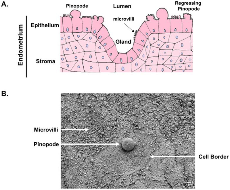
Successful embryo implantation is a complex and highly regulated process involving precise synchronization between the fetal-derived trophoblast cells and maternal uterine luminal epithelium. Multiple endocrine-driven factors are important for controlling the timely receptivity of the uterus, and this complexity underscores implantation failure as a major cause of recurrent infertility associated with assisted reproductive technologies. One particular cellular structure often hypothesized to promote receptivity is the pinopode or uterodome - a hormonally regulated, large cellular protrusion on the uterine epithelial surface. Recent clinical studies associate pinopodes with favorable fertility outcomes in women, and because they are directly linked to an increase in progesterone levels, the potential utility of these hormone-regulated cell biological structures in predicting or improving implantation in a clinical setting holds promise. In this review, we aim to generate interest in pinopodes from the broader cell biology and endocrinology communities, re-examine methodologies in pinopode research, and identify priorities for future investigation of pinopode structure and function in women's reproductive health.
Keywords: Endometrial receptivity; Implantation; Pinopodes; Progesterone; Uterodomes.
Copyright © 2019 Elsevier B.V. All rights reserved.
Conflict of interest statement
Conflict of Interest:
The authors have declared that no conflict of interest exists.
Figures
References
-
- Acosta AA, Elberger L, Borghi M, Calamera JC, Chemes H, Doncel GF, Kliman H, Lema B, Lustig L, and Papier S (2000). Endometrial dating and determination of the window of implantation in healthy fertile women. Fertil Steril 73, 788–798. - PubMed
-
- Adams SM, Gayer N, Hosie MJ, and Murphy CR (2002). Human uterodomes (pinopods) do not display pinocytotic function. Hum Reprod 17, 1980–1986. - PubMed
-
- Aghajanova L, Stavreus-Evers A, Nikas Y, Hovatta O, and Landgren BM (2003). Coexpression of pinopodes and leukemia inhibitory factor, as well as its receptor, in human endometrium. Fertil Steril 79 Suppl 1, 808–814. - PubMed
-
- Aplin JD (1997). Adhesion molecules in implantation. Rev Reprod 2, 84–93. - PubMed
Publication types
MeSH terms
Substances
Grants and funding
LinkOut - more resources
Full Text Sources
Other Literature Sources
