. 2020 Jan 22;88(2):e00759-19.
doi: 10.1128/IAI.00759-19. Print 2020 Jan 22.

Dendritic Cells Targeting Lactobacillus plantarum Strain NC8 with a Surface-Displayed Single-Chain Variable Fragment of CD11c Induce an Antigen-Specific Protective Cellular Immune Response

Affiliations

Dendritic Cells Targeting Lactobacillus plantarum Strain NC8 with a Surface-Displayed Single-Chain Variable Fragment of CD11c Induce an Antigen-Specific Protective Cellular Immune Response

Jing Liu et al. Infect Immun. .

Abstract

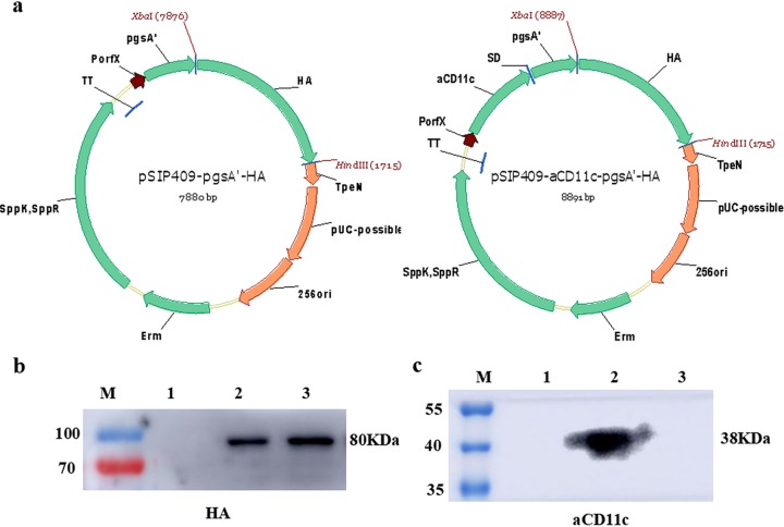
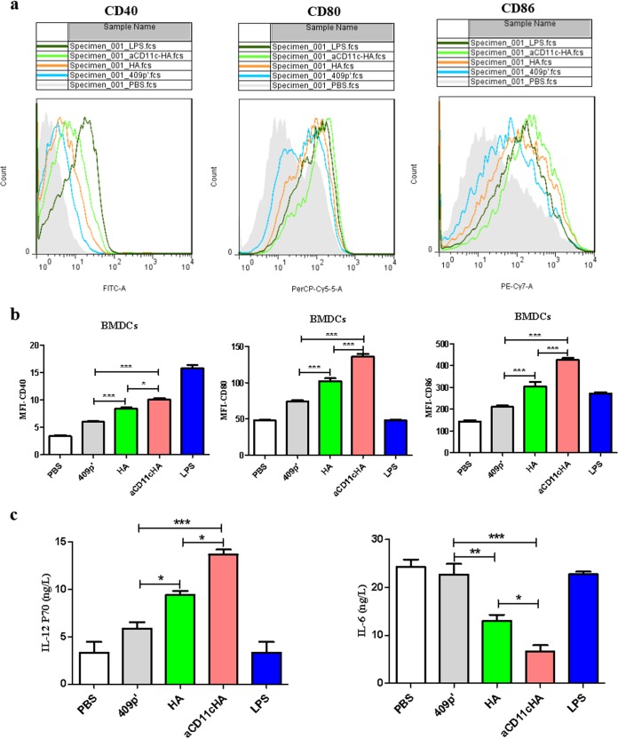
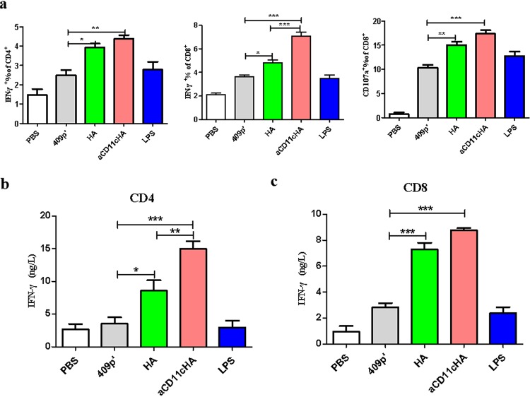
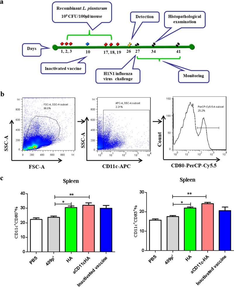
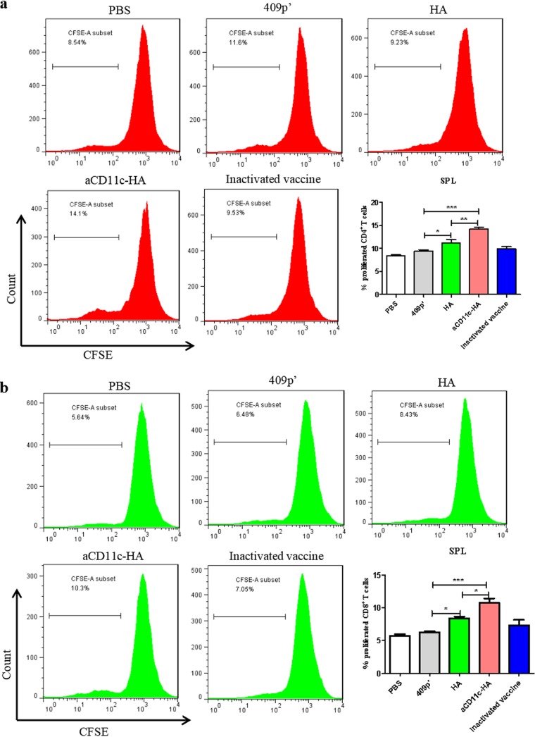
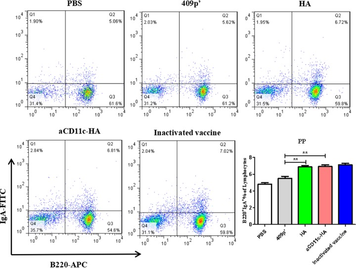
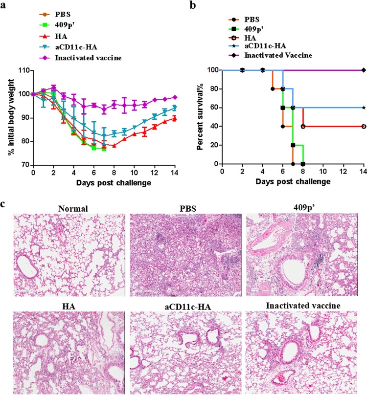
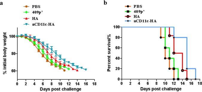
Influenza A virus (H1N1) is an acute, highly contagious respiratory virus. The use of lactic acid bacteria (LAB) to deliver mucosal vaccines against influenza virus infection is a research hot spot. In this study, two recombinant Lactobacillus plantarum strains expressing hemagglutinin (HA) alone or coexpressing aCD11c-HA to target HA protein to dendritic cells (DCs) by fusion to an anti-CD11c single-chain antibody (aCD11c) were constructed. The activation of bone marrow dendritic cells (BMDCs) by recombinant strains and the interaction of activated BMDCs and sorted CD4+ or CD8+ T cells were evaluated through flow cytometry in vitro, and cellular supernatants were assessed by using an enzyme-linked immunosorbent assay kit. The results demonstrated that, compared to the HA strain, the aCD11c-HA strain significantly increased the activation of BMDCs and increased the production of CD4+ gamma interferon-positive (IFN-γ+) T cells, CD8+ IFN-γ+ T cells, and IFN-γ in the cell culture supernatant in vitro Consistent with these results, the aCD11c-HA strain clearly increased the activation and maturation of DCs, the HA-specific responses of CD4+ IFN-γ+ T cells, CD8+ IFN-γ+ T cells, and CD8+ CD107a+ T cells, and the proliferation of T cells in the spleen, finally increasing the levels of specific antibodies and neutralizing antibodies in mice. In addition, the protection of immunized mice was observed after viral infection, as evidenced by improved weight loss, survival, and lung pathology. The adoptive transfer of CD8+ T cells from the aCD11c-HA mice to NOD/Lt-SCID mice resulted in a certain level of protection after influenza virus infection, highlighting the efficacy of the aCD11c targeting strategy.

Keywords: Lactobacillus plantarum; cellular immune response; dendritic cell targeting.


Figures

FIG 1
FIG 1
Structural diagrams of pSIP409-pgsA′-HA and pSIP409-aCD11c-pgsA′-HA plasmids and detection of HA and aCD11c expression. (a) pSIP409-pgsA′-HA and pSIP409-aCD11c-pgsA′-HA were constructed as described in the text. pgsA′, anchoring sequence; HA, heterologous protein (hemagglutinin); aCD11c, scFv-CD11c. The expression of HA (b) and aCD11c (c) in L. plantarum was measured by Western blotting. HA and His were used as primary antibodies in panels b and c, respectively. M, prestained marker; lane 1, 409p′ strain; lane 2, aCD11c-HA strain; lane 3, HA strain.
FIG 2
FIG 2
The aCD11c-HA strain promotes the activation of BMDCs and the secretion of cytokines. (a) The flow peak diagrams from PBS, 409p′, HA, and aCD11c-HA groups are displayed. LPS was used as a positive control. (b) MFIs of CD40, CD80, and CD86 from each group analyzed by FCM. (c) Concentrations of IL-12P70 and IL-6 in cell culture supernatants measured by ELISA. The statistical significance was calculated using one-way ANOVA (*, P < 0.05; **, P < 0.01; ***, P < 0.001).
FIG 3
FIG 3
T cell activation in vitro. Sorted CD4+ T cells and CD8+ T cells were cocultured with the differentiated BMDCs for 48 h. (a) IFN-γ-producing CD4+ T cells or IFN-γ-, CD107a-producing CD8+ T cells evaluated by FCM. The cell culture supernatants were used to detect the secretion of IFN-γ from CD4+ (b) and CD8+ T cells (c) by ELISA. The statistical significance was calculated via one-way ANOVA (*, P < 0.05; **, P < 0.01; ***, P < 0.001).
FIG 4
FIG 4
Immunization protocol and the in vivo differentiation and maturation of DCs. (a) Protocol for mouse immunization. (b and c) Proportions of CD11c-, CD80-, and CD83-expressing splenocytes, as evaluated by FCM. The statistical significance was calculated by using one-way ANOVA (*, P < 0.05; **, P < 0.01).
FIG 5
FIG 5
T cell immune responses in vivo. (a) HA-specific cytokine secretion from splenocytes and MLNs was measured by FCM after oral immunization. The percentages of HA-specific CD4+ IFN-γ+ T cells, CD8+ IFN-γ+ T cells, and CD8+CD107a+ T cells from the spleen (b) and MLN (c) were detected by using FCM. The mean values ± the SEM of three independent experiments are shown. The statistical significance was calculated by using one-way ANOVA (*, P < 0.05; **, P < 0.01; ***, P < 0.001).
FIG 6
FIG 6
T cell proliferation assay. HA protein was used to stimulate the proliferation of mouse splenic T cells. The percentages of CD4+ T cell (a) and CD8+ T cell (b) proliferation were evaluated in CFSE (carboxyfluorescein succinimidyl ester)-stained splenocytes by FCM. The statistical significance was calculated by one-way ANOVA (*, P < 0.05; **, P < 0.01; ***, P < 0.001).
FIG 7
FIG 7
Percentages of B220+ IgA+ B cells in PPs. The flow scatter diagrams of each group are displayed, and the percentages of B220+ IgA+ B cells in the PPs were evaluated. The statistical significance was calculated using a one-way ANOVA (**, P < 0.01).
FIG 8
FIG 8
Determination of HA-specific antibodies and neutralizing antibodies. The HA-specific serum IgG titer (a) and the fecal (b) and BALF (c) IgA antibody titers were detected by indirect ELISA. Purified HA protein was used as the plate-coating antigen. Endpoint titers (log2) were calculated in accordance with the dilution of the serum, feces, and BALF by indirect ELISA. (d) Neutralizing antibody levels were detected in 10-day-old embryonated SPF eggs. Neutralizing antibody titers (log10) were calculated by the Reed and Muench method (39). The statistical significance was calculated using one-way ANOVA (**, P < 0.01; ***, P < 0.001).
FIG 9
FIG 9
Protection against influenza virus challenge. Weight loss (a) and survival (b) were recorded for 2 weeks. The weight loss and mortality data are presented as the means ± the SEM of triplicate tests (n = 5 mice per group). (c) Lung samples from different influenza virus-infected groups were acquired, fixed, and embedded in paraffin. Sections were stained with H&E (magnification, ×200). Scale bar, 200 μm.
FIG 10
FIG 10
Protective effect of CD8+ T cells transferred from aCD11c-HA mice to NOD/Lt-SCID mice. (a) The weight loss of the mice after H1N1 influenza virus infection was detected every day until day 16. (b) The survival rates of the NOD/Lt-SCID mice were observed after virus infection. The weight loss and mortality data are shown as means ± the SEM of triplicate tests (n = 5 mice per group).

References

    1. Mohebbi A, Fotouhi F, Jamali A, Yaghobi R, Farahmand B, Mohebbi R. 2019. Molecular epidemiology of the hemagglutinin gene of prevalent influenza virus A/H1N1/pdm09 among patient in Iran. Virus Res 259:38–45. doi:10.1016/j.virusres.2018.10.001. - DOI - PubMed
    1. Yang WT, Yang GL, Wang Q, Huang HB, Jiang YL, Shi CW, Wang JZ, Huang KY, Jin YB, Wang CF. 2017. Protective efficacy of Fc targeting conserved influenza virus M2e antigen expressed by Lactobacillus plantarum. Antiviral Res 138:9–21. doi:10.1016/j.antiviral.2016.11.025. - DOI - PubMed
    1. Fraser C, Donnelly CA, Cauchemez S, Hanage WP, Van Kerkhove MD, Hollingsworth TD, Griffin J, Baggaley RF, Jenkins HE, Lyons EJ, Jombart T, Hinsley WR, Grassly NC, Balloux F, Ghani AC, Ferguson NM, Rambaut A, Pybus OG, Lopez-Gatell H, Alpuche-Aranda CM, Chapela IB, Zavala EP, Guevara DM, Checchi F, Garcia E, Hugonnet S, Roth C, The WHO Rapid Pandemic Assessment Collaboration . 2009. Pandemic potential of a strain of influenza A (H1N1): early findings. Science 324:1557–1561. doi:10.1126/science.1176062. - DOI - PMC - PubMed
    1. Deng L, Roose K, Job ER, De Rycke R, Van Hamme E, Goncalves A, Parthoens E, Cicchelero L, Sanders N, Fiers W, Saelens X. 2017. Oral delivery of Escherichia coli persistently infected with M2e-displaying bacteriophages partially protects against influenza A virus. J Control Release 264:55–65. doi:10.1016/j.jconrel.2017.08.020. - DOI - PubMed
    1. Te Velthuis AJ, Fodor E. 2016. Influenza virus RNA polymerase: insights into the mechanisms of viral RNA synthesis. Nat Rev Microbiol 14:479–493. doi:10.1038/nrmicro.2016.87. - DOI - PMC - PubMed

Publication types

MeSH terms