. 2019 Dec 27;294(52):19978-19987.
doi: 10.1074/jbc.RA119.011435. Epub 2019 Nov 18.

Structural and biochemical studies of the glucuronoyl esterase Ot CE15A illuminate its interaction with lignocellulosic components

Affiliations

Structural and biochemical studies of the glucuronoyl esterase Ot CE15A illuminate its interaction with lignocellulosic components

Scott Mazurkewich et al. J Biol Chem. .

Abstract

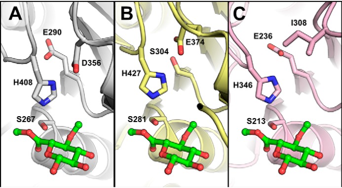
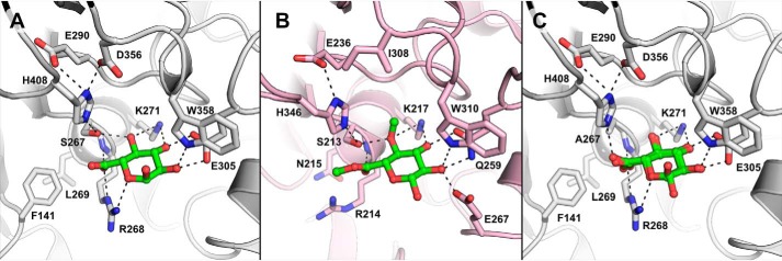
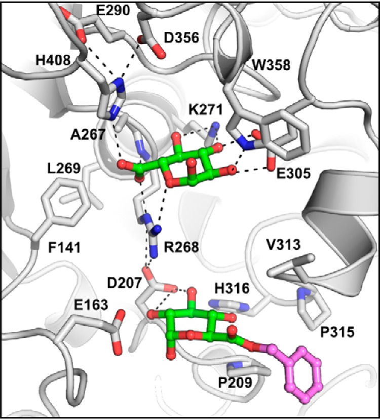
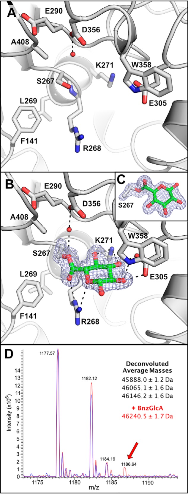
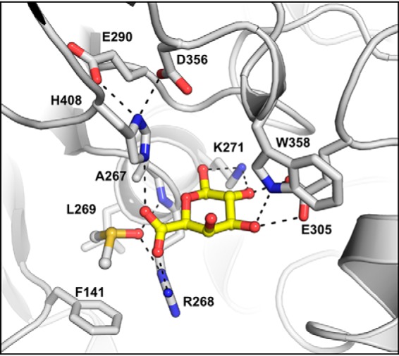
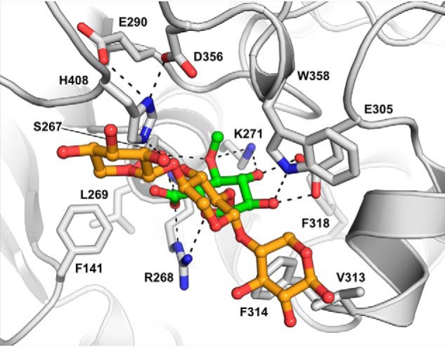
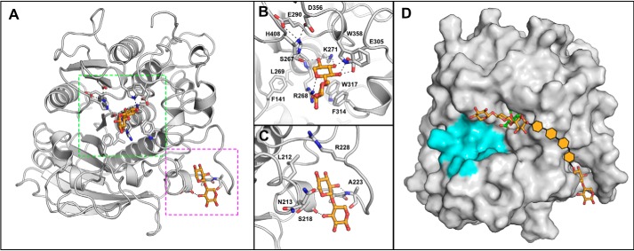
Glucuronoyl esterases (GEs) catalyze the cleavage of ester linkages between lignin and glucuronic acid moieties on glucuronoxylan in plant biomass. As such, GEs represent promising biochemical tools in industrial processing of these recalcitrant resources. However, details on how GEs interact and catalyze degradation of their natural substrates are sparse, calling for thorough enzyme structure-function studies. Presented here is a structural and mechanistic investigation of the bacterial GE OtCE15A. GEs belong to the carbohydrate esterase family 15 (CE15), which is in turn part of the larger α/β-hydrolase superfamily. GEs contain a Ser-His-Asp/Glu catalytic triad, but the location of the catalytic acid in GEs has been shown to be variable, and OtCE15A possesses two putative catalytic acidic residues in the active site. Through site-directed mutagenesis, we demonstrate that these residues are functionally redundant, possibly indicating the evolutionary route toward new functionalities within the family. Structures determined with glucuronate, in both native and covalently bound intermediate states, and galacturonate provide insights into the catalytic mechanism of CE15. A structure of OtCE15A with the glucuronoxylooligosaccharide 23-(4-O-methyl-α-d-glucuronyl)-xylotriose (commonly referred to as XUX) shows that the enzyme can indeed interact with polysaccharides from the plant cell wall, and an additional structure with the disaccharide xylobiose revealed a surface binding site that could possibly indicate a recognition mechanism for long glucuronoxylan chains. Collectively, the results indicate that OtCE15A, and likely most of the CE15 family, can utilize esters of glucuronoxylooligosaccharides and support the proposal that these enzymes work on lignin-carbohydrate complexes in plant biomass.

Keywords: alpha/beta-hydrolase; biomass; carbohydrate processing; carbohydrate-active enzymes; catalytic triad; cell wall; enzyme kinetics; enzyme mutation; enzyme structure; glucuronoxylan; glucuronoyl esterase; lignin-carbohydrate complexes.


Conflict of interest statement

The authors declare that they have no conflicts of interest with the contents of this article

Figures

Figure 1.
Figure 1.
Catalytic residues of OtCE15A and other CE15 members. Comparison of the catalytic triads of OtCE15A (PDB code 6GS0; A), TtCE15A from T. turnerae (PDB code 6HSW; B), and StGE2 from Thermothelomyces thermophila (previously Sporotrichum thermophile; PDB code 4G4G; C) shows alternate positions of the catalytic acid residue in TtCE15A and StGE2, whereas an acidic residue is present in both locations in OtCE15A. The methyl ester of 4-O-methyl glucuronoate determined in the structure of StGE2 (PDB code 4G4J) is shown in green sticks from structural alignment of the CE15 proteins.
Figure 2.
Figure 2.
Glucuronate bound to OtCE15A. Binding of glucuronate to the WT OtCE15A (A; PDB code 6SYR) and S267A OtCE15A variant (C; showing both anomers of the carbohydrate; PDB code 6SYV) is similar to that of the methyl ester of 4-O-methyl glucuoronoate determined in the structure of StGE2 from T. thermophila (B; previously S. thermophile; PDB code 4G4G with the ligand taken from 4G4J by structural alignment). Notably, the side chain of the conserved active-site arginine is found rotated toward, and interacts with, the glucuronate molecule in the OtCE15A product ligated structures differently than in the StGE2 substrate ligated structure.
Figure 3.
Figure 3.
Benzyl glucuronoate trapped on the surface of the S267A OtCE15A variant (PDB code 6T0E). Presumably, the cleavage rate was too fast to capture the Michaelis–Menten complex with benzyl glucuronoate over the short crystal soaking period (10 s) but allowed capture of the uncleaved substrate along the edge of the active site cleft ∼10 Å from the catalytic center (glucuronate portion in green and benzyl portion in magenta).
Figure 4.
Figure 4.
Trapping the glucuronate covalent intermediate in the OtCE15A H408A variant. Shown is the OtCE15A H408A variant in the absence (A; PDB code 6SZ0) and presence of benzyl glucuronoate (B; PDB code 6SZ4). Presumably, the acylation rate was too fast to capture the Michaelis–Menten complex with benzyl glucuronoate over the short crystal soaking period (5 s) but allowed capture of the acyl-enzyme intermediate with the glucuronate moiety covalently linked to the catalytic nucleophile Ser-267. In both structures, a water molecule, hydrogen-bonded to Asp-356, fills the void left from substitution of the catalytic histidine. The covalent serine-glucuronoyl adduct is shown with the density from an omit map, at 4σ, created in Phenix (39) by omitting GlcA and the Cα, Cβ, and Oγ of Ser-267. C, an alternate orientation of the serine-glucuronoyl adduct showing the linkage. D, mass spectrum of the OtCE15A H408A variant in the absence (blue) and presence of benzyl glucuronate (red) leads to the production of a new mass consistent with the glucuronate covalent intermediate.
Figure 5.
Figure 5.
Galacturonate bound to the OtCE15A S267A variant (PDB code 6SZO). Presumably, the cleavage rate was too fast to capture a Michaelis–Menten complex with methyl galacturonoate over the crystal soaking period (60 s) but allowed capture of the galacturonate product in the active site. The galacturonate is shown in yellow sticks, and a DMSO molecule, used as a solvent for the substrate, present in the oxyanion hole is shown in sticks.
Figure 6.
Figure 6.
OtCE15A in complex with the glucuronoxylan oligosaccharide XUX (PDB code 6T0I). The oligosaccharide, produced from beech xylan as described under “Experimental procedures,” is shown with the 4-O-methyl-α-d-glucuronate moiety in green sticks and the xylotriose moiety in orange sticks.
Figure 7.
Figure 7.
OtCE15A in complex with xylobiose. Shown is the overall structure of OtCE15A, showing the two xylobiose-binding sites (PDB code 6SYU) highlighted in green and magenta (A) for the active site (B) and secondary site (C), respectively. The secondary site is ∼25 Å from the active site. D, a proposal of how the secondary xylobiose site could connect to the XUX found in the active site. Potential xylose moieties connecting the xylobiose observed in the second site to the XUX molecule observed in the active site are shown by orange hexagons. The region of the protein proposed to interact with lignin is colored in cyan.

References

    1. Biely P. (2016) Microbial glucuronoyl esterases: 10 years after discovery. Appl. Environ. Microbiol. 82, 7014–7018 10.1128/AEM.02396-16 - DOI - PMC - PubMed
    1. Spániková S., and Biely P. (2006) Glucuronoyl esterase—novel carbohydrate esterase produced by Schizophyllum commune. FEBS Lett. 580, 4597–4601 10.1016/j.febslet.2006.07.033 - DOI - PubMed
    1. Lombard V., Golaconda Ramulu H., Drula E., Coutinho P. M., and Henrissat B. (2014) The carbohydrate-active enzymes database (CAZy) in 2013. Nucleic Acids Res. 42, D490–D495 10.1093/nar/gkt1178 - DOI - PMC - PubMed
    1. Arnling Bååth J., Giummarella N., Klaubauf S., Lawoko M., and Olsson L. (2016) A glucuronoyl esterase from Acremonium alcalophilum cleaves native lignin-carbohydrate ester bonds. FEBS Lett. 590, 2611–2618 10.1002/1873-3468.12290 - DOI - PubMed
    1. Mosbech C., Holck J., Meyer A. S., and Agger J. W. (2018) The natural catalytic function of CuGE glucuronoyl esterase in hydrolysis of genuine lignin-carbohydrate complexes from birch. Biotechnol. Biofuels 11, 71 10.1186/s13068-018-1075-2 - DOI - PMC - PubMed

Publication types

MeSH terms

Supplementary concepts