. 2020 Jan;5(1):126-140.
doi: 10.1038/s41564-019-0588-1. Epub 2019 Nov 18.

Cultivation and functional characterization of 79 planctomycetes uncovers their unique biology

Affiliations

Cultivation and functional characterization of 79 planctomycetes uncovers their unique biology

Sandra Wiegand et al. Nat Microbiol. 2020 Jan.

Abstract

When it comes to the discovery and analysis of yet uncharted bacterial traits, pure cultures are essential as only these allow detailed morphological and physiological characterization as well as genetic manipulation. However, microbiologists are struggling to isolate and maintain the majority of bacterial strains, as mimicking their native environmental niches adequately can be a challenging task. Here, we report the diversity-driven cultivation, characterization and genome sequencing of 79 bacterial strains from all major taxonomic clades of the conspicuous bacterial phylum Planctomycetes. The samples were derived from different aquatic environments but close relatives could be isolated from geographically distinct regions and structurally diverse habitats, implying that 'everything is everywhere'. With the discovery of lateral budding in 'Kolteria novifilia' and the capability of the members of the Saltatorellus clade to divide by binary fission as well as budding, we identified previously unknown modes of bacterial cell division. Alongside unobserved aspects of cell signalling and small-molecule production, our findings demonstrate that exploration beyond the well-established model organisms has the potential to increase our knowledge of bacterial diversity. We illustrate how 'microbial dark matter' can be accessed by cultivation techniques, expanding the organismic background for small-molecule research and drug-target detection.


Figures

Figure 1. |
Figure 1. |. Sampling the phylum Planctomycetes.
A. Phylogeny and environmental occurrences of the planctomycetal isolates. Maximum-likelihood phylogenetic tree of 9,002 non-redundant planctomycetal 16S rRNA gene sequences, including the isolates (red) and MAGs (yellow) reported in this study as well as resequenced strains (green; Supplementary Data 1). The size of the collapsed clades corresponds to the number of sequences they comprise (between 10 and 104); clades with fewer than ten sequences have been condensed to a single line. The occurrence of the 16S rRNA genes of reported cultures in publicly available amplicon datasets from the NCBI Sequence Read Archive was analyzed with a 99% sequence-identity threshold designed to identify identical and closely related strains. The detailed distribution of the respective hits is illustrated as a corresponding pie chart for the cases where >10 hits against planctomycetes (total) were found to comprise >0.02% of a given dataset, of which >0.005% were strain specific. The number of positively screened datasets is illustrated by the respective size of the plotted pie charts (between 11 and 3,806 hits in the analyzed studies). The corresponding sectors are color-coded according to the habitat categories of the respective dataset. The outer border of each pie chart is color-coded according to the origin of the isolate itself. B. Diversity-driven sampling. The samples for this study were obtained from a variety of aquatic locations, mostly throughout Europe, and include iron-hydroxide deposits (D); microbial mats from methane seeps in the Black Sea (K); underwater fluid discharges (gas and water) and hot lakes in the Tyrrhenian Sea around Panarea island, Italy (P); kelp surfaces at Monterey Bay, California (M); surfaces of different macroalgae and jellyfish species at Heligoland island, Germany (H); seaweed meadows and calcareous sponges of the Mediterranean Sea (C); freshwater sponges from Lake Constance (L) and Lake Salzgitter (Z), Germany; beach sediment and algae at El Arenal, Mallorca island, Spain (E); offshore seawater of Costa Brava, Spain (V); freshwater from a meromictic lake in Bergen, Norway (N); a cyanobacterial bloom in the Stadtgraben pond in Wolfenbüttel, Germany; a seawater ornamental aquarium; wood, polyethylene and polystyrene baits deposited at Warnow River estuary, Rostock, Germany and costal Baltic Sea spots, Heiligendamm, Germany (B) and hypoxic and anaerobic bioreactors (A).
Figure 2.
Figure 2.. Current diversity of the planctomycetal phylum.
Core information assembled in this study briefly summarizing all other figures and additional data. The centrepiece of the figure is formed by a MLSA-based phylogenetic tree of all of the isolates and MAGs from this study and from the resequenced or publicly available genomes of previously described strains, enrichment cultures and metagenomic samples (a total of 150). Details on the tree as well as the rooting and branching probabilities can be found in Supplementary Fig.1, Supplementary Data 2 and Methods; details on the genomes reported in this study are given in Supplementary Table 1. The focal point is encompassed by Circles 1 and 2, which highlight the different subclades that constitute the planctomycetal phylum and the current knowledge on the distribution of different cell-division strategies throughout it. The first inner circles hold information on the sampling sites and the environmental conditions there (Circles 6 and 7) as well as the main properties of the gathered genomes (Circle 5). They are followed by Circles 8–11, illustrating the number of identified putative secondary metabolite BGCs, giant genes, two-component systems and ECF σ factors. The innermost Circles 12–19 provide an overview of the presence or absence of the canonical genes involved in peptidoglycan biosynthesis, cytoskeleton formation and cell division.
Figure 3.
Figure 3.. Planctomycetal cell division.
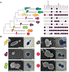
A. Collapsed phylogenetic tree of the planctomycetal subclades with their respective modes of cell division and the presence (filled circles) or absence (empty circles) of canonical cell-division genes (see also Supplementary Fig. 7). For an uncollapsed version of the tree please refer to Fig. 2 or Supplementary Fig. 1. The genes mreB, ftsI and ftsW were deleted in P. limnophila (marked by a red X) without observing a phenotype. B. Most of the known planctomycetes, such as the pear-shaped P. limnophila ((i) and (ii)), divide by polar budding. Budding was indeed frequently observed among our isolates, not only in cells with an elongated shape but also in coccoid cells such as ‘Aeolisphaera plasticia’ ElP ((iii) and (iv)). In contrast, members of the class Phycisphaerae – for example, ‘Mucisphaera calidilacus’ Pan265 ((v) and (vi)) – divide by binary fission. A previously unseen mode of cell division was observed for ‘Kolteria novifilia’ Pan216, which divides by lateral budding ((vii) and (viii); Supplementary Movie 1). Members of the Saltatorellus clade on the other hand have the ability to perform budding ((ix) and (x); Poly30) as well as binary fission ((xi) and (xii); Poly30; Supplementary Movie 2), even in the same culture. The first and the third columns of images are scanning electron micrographs; the second and fourth columns contain phase-contrast light-microscopy images. Scale bars, 1 μm. The selected micrographs originate from at least two independent experiments; more than 100 cells were analyzed and the best representative image was chosen.
Figure 4.
Figure 4.. Signaling in Planctomycetes.
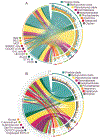
A. Signal transduction systems. A total of 26,487 components related to signal-transduction systems were identified in the 150 analyzed genomes (Supplementary Table 6). The analyzed planctomycetal genomes are depicted on the right-hand side of the circular plot. Each column of the stacked-column chart represents one strain and the different stacks of the columns outline the different signal-transduction-system components found in each genome (see the lower-left legend). The colors of the overlying arcs and the underlying links suggest the planctomycetal clade that contains the respective strain (see the upper-right legend). In addition, the links connect each strain to the category of the identified signal-transduction-system components on the left-hand side of the plot. Two-component signal-transduction systems: HisK, two-component sensor histidine kinases and REC, response regulators. Chemotaxis: MCPs. c-di-GMP hydrolysis: GGDEF, diguanylate cyclases; EAL, EAL-domain containing c-di-GMP phosphodiesterases; HD-GyP, HD-GyP-domain containing c-di-GMP phosphodiesterases. Ser/Thr/Tyr protein kinases and phosphoprotein phosphatases: STyK, Ser/Thr/Tyr protein kinases; PP2C, PP2C-type protein phosphatases. ACyc3, class III adenylate/guanylate cyclases. B. Extracytoplasmic function σ factors. A total of 5,966 ECF σ factors were identified in all 150 analyzed genomes (Supplementary Table 7). A subsequent similarity clustering found them to form 4,694 clusters, which are indicated on the left-hand side of the circular plot. The outer histogram marks the total number of identified ECF σ factors per cluster and the underlying curves/arcs define the groups of the ECF σ factors (see the lower-left legend). The right-hand side of the circular plot describes the analyzed planctomycetal genomes. Each column of the stacked-column chart represents one strain and the different stacks of the columns outline the ECF σ factors found in each genome (see the lower-left legend). The colors of the overlying arcs and the underlying links suggest the planctomycetal clade that encompasses the respective strain (see the upper-right legend). Finally, the links point to the ECF σ factor groups that the ECF σ factors of each strain belong to.
Figure 5.
Figure 5.. Secondary metabolite biosynthetic gene clusters (BGCs).
A total of 1,104 BGCs with potential involvement in the production of secondary metabolites were identified in all 196 analyzed genomes (Supplementary Table 10). Many of these BGCs fall into 162 clusters, but 359 are unique according to the applied parameters. These 521 BGC groups are provided on the left-hand side of the chart, with the histogram marking the total number of identified BGCs per group. The underlying arcs define the BGC functional category (see the lower-left legend) of the group. On the right-hand side of the chart, each column of the stacked column chart represents one strain and the different stacks of the columns sum up the BGCs found in each genome (see the lower-left legend). The colors of the overlying arcs and the connected links indicate the planctomycetal clade that encompasses the respective strain (see the upper-right legend). The links connect the hits for each strain to the corresponding BGC group. A cluster analysis of the identified BGCs with previously described BGCs is shown in Supplementary Fig. 45 and provides the relatedness of the different BGCs. NRPS, non-ribosomal peptide synthases.

References

    1. Wiegand S, Jogler M & Jogler C On the maverick Planctomycetes. FEMS Microbiol. Rev 42, 739–760 (2018). - PubMed
    1. Wagner M & Horn M The Planctomycetes, Verrucomicrobia, Chlamydiae and sister phyla comprise a superphylum with biotechnological and medical relevance. Curr. Opin. Biotechnol 17, 241–249 (2006). - PubMed
    1. Peeters SH & van Niftrik L Trending topics and open questions in anaerobic ammonium oxidation. Curr. Opin. Chem. Biol 49, 45–52 (2018). - PubMed
    1. Jeske O et al. Developing techniques for the utilization of Planctomycetes as producers of bioactive molecules. Front. Microbiol 7, 1242 (2016). - PMC - PubMed
    1. van Teeseling MCF et al. Anammox Planctomycetes have a peptidoglycan cell wall. Nat. Commun 6, 6878 (2015). - PMC - PubMed

Publication types

Substances