. 2019 Sep 30;50(1):31-39.
doi: 10.1002/jmd2.12060. eCollection 2019 Nov.

A mutation in mannose-phosphate-dolichol utilization defect 1 reveals clinical symptoms of congenital disorders of glycosylation type I and dystroglycanopathy

Affiliations

A mutation in mannose-phosphate-dolichol utilization defect 1 reveals clinical symptoms of congenital disorders of glycosylation type I and dystroglycanopathy

Walinka van Tol et al. JIMD Rep. .

Abstract

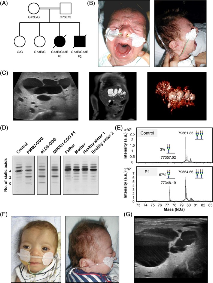
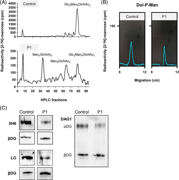
Congenital disorders of glycosylation type I (CDG-I) are inborn errors of metabolism, generally characterized by multisystem clinical manifestations, including developmental delay, hepatopathy, hypotonia, and skin, skeletal, and neurological abnormalities. Among others, dolichol-phosphate-mannose (DPM) is the mannose donor for N-glycosylation as well as O-mannosylation. DOLK-CDG, DPM1-CDG, DPM2-CDG, and DPM3-CDG are defects in the DPM synthesis showing both CDG-I abnormalities and reduced O-mannosylation of alpha-dystroglycan (αDG), which leads to muscular dystrophy-dystroglycanopathy. Mannose-phosphate-dolichol utilization defect 1 (MPDU1) plays a role in the utilization of DPM. Here, we report two MPDU1-CDG patients without skin involvement, but with massive dilatation of the biliary duct system and dystroglycanopathy characteristics including hypotonia, elevated creatine kinase, dilated cardiomyopathy, buphthalmos, and congenital glaucoma. Biochemical analyses revealed elevated disialotransferrin in serum, and analyses in fibroblasts showed shortened lipid linked oligosaccharides and DPM, and reduced O-mannosylation of αDG. Thus, MPDU1-CDG can be added to the list of disorders with overlapping biochemical and clinical abnormalities of CDG-I and dystroglycanopathy.

Synopsis: Mannose-phosphate-dolichol utilization defect 1 patients can have overlapping biochemical and clinical abnormalities of congenital disorders of glycosylation type I and dystroglycanopathy.

Keywords: MPDU1‐CDG; congenital disorders of glycosylation; dolichol‐phosphate‐mannose; dystroglycanopathy.


Conflict of interest statement

The authors declare that they have no conflict of interest.

Figures

Figure 1
Figure 1
Family pedigree and clinical images of affected patients. A, Family pedigree of MPDU1 patients (P1 = patient 1, P2 = patient 2). B, Front and side facial view of patient 1. C, Abdominal sonography (left), magnetic resonance imaging (middle), and 3D image from magnetic resonance cholangiopancreatography (right) of patient 1 showing a vast dilatation of the complete intrahepatic biliary duct system. D, Serum transferrin isoelectric focusing (TIEF) analysis of P1, her parents and two healthy sisters. E, ESI‐MS of serum transferrin. Disialotransferrin levels are expressed as a percentage of tetrasialotransferrin. F, Front and side facial view of patient 2 at 4 months of age. G, Abdominal sonography of patient 2, showing dilatation of the intrahepatic biliary duct. CDG, congenital disorders of glycosylation; ESI‐MS, electrospray ionization mass spectrometry; MPDU1, mannose‐phosphate‐dolichol utilization defect 1
Figure 2
Figure 2
CDG diagnostics and biochemical analyses of MPDU1‐CDG P1. A, HPLC analysis of LLO from fibroblasts of patient 1 (P1) and a control revealed the accumulation of the shortened dolichol‐linked oligosaccharides Man5GlcNAc2 and Man9GlcNAc2. B, Thin‐layer chromatography (TLC) analysis of hydrophobic LLO extracts further revealed that dolichol‐phosphate‐mannose (Dol‐P‐Man) is synthesized in patient 1 fibroblasts. C, Analysis of O‐mannosylated αDG in P1 and control fibroblasts. IIH6 and laminin (laminin overlay, LO) only bind to fully functional O‐mannosyl glycans of αDG. DAG1 binds to the core of the dystroglycan protein, showing expression of αDG and βDG proteins. CDG, congenital disorders of glycosylation; HPLC, High‐performance liquid chromatography; LLO, lipid linked oligosaccharide; MPDU1, mannose‐phosphate‐dolichol utilization defect 1

References

    1. Jaeken J, Péanne R. What is new in CDG? J Inherit Metab Dis. 2017;40:621‐625. - PubMed
    1. Barone R, Aiello C, Race V, et al. DPM2‐CDG: a muscular dystrophy‐dystroglycanopathy syndrome with severe epilepsy. Ann Neurol. 2012;72:550‐558. - PubMed
    1. Lefeber DJ, de Brouwer APM, Morava E, et al. Autosomal recessive dilated cardiomyopathy due to DOLK mutations results from abnormal dystroglycan O‐mannosylation. PLoS Genet. 2011;7:e1002427. - PMC - PubMed
    1. Lefeber DJ, Schönberger J, Morava E, et al. Deficiency of Dol‐P‐Man synthase subunit DPM3 bridges the congenital disorders of glycosylation with the dystroglycanopathies. Am J Hum Genet. 2009;85:76‐86. - PMC - PubMed
    1. Yang AC, Ng BG, Moore SA, et al. Congenital disorder of glycosylation due to DPM1 mutations presenting with dystroglycanopathy‐type congenital muscular dystrophy. Mol Genet Metab. 2013;110:345‐351. - PMC - PubMed