

Surgical Excision with Rotational Flap Reconstruction for Basosquamous Cell Carcinoma in Xeroderma Pigmentosa: A Case Report
- PMID: 31742046
- PMCID: PMC6848524
- DOI: 10.1007/s12070-018-1508-z
Surgical Excision with Rotational Flap Reconstruction for Basosquamous Cell Carcinoma in Xeroderma Pigmentosa: A Case Report
Abstract
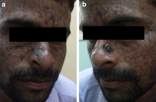
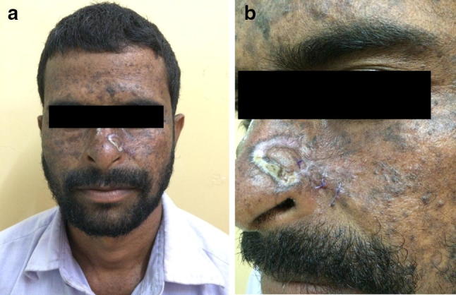
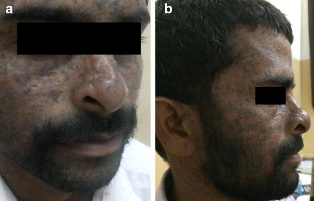
Xeroderma pigmentosa (XP) is a rare autosomal recessive disorder which is characterized by a defect in nucleotide excision repair of DNA following exposure to UV radiation. This leads to hypersensitivity to sunlight causing pigmented skin lesions, photophobia and a 1000-fold increase in risk of developing cutaneous malignancies like basal cell carcinomas, squamous cell carcinomas and melanomas of head and neck. We present an interesting case of a 28 year old man with basosquamous cell carcinoma secondary to XP treated successfully with surgical excision and reconstruction with rotational flap technique.
Keywords: Basal cell carcinoma; Rotational flap; Xeroderma pigmentosa.
© Association of Otolaryngologists of India 2018.
Figures


Similar articles
-
Cutaneous malignancies in xeroderma pigmentosum: earlier management improves survival.Indian J Otolaryngol Head Neck Surg. 2013 Apr;65(2):162-7. doi: 10.1007/s12070-012-0614-6. Epub 2013 Jan 5. Indian J Otolaryngol Head Neck Surg. 2013. PMID: 24427558 Free PMC article.
-
Xeroderma Pigmentosum with Simultaneous Cutaneous and Ocular Squamous Cell Carcinoma.Clin Cosmet Investig Dermatol. 2022 Feb 2;15:157-161. doi: 10.2147/CCID.S348771. eCollection 2022. Clin Cosmet Investig Dermatol. 2022. PMID: 35140492 Free PMC article.
-
Xeroderma pigmentosa associated with spindle cell carcinoma of lung: a case report.Ann Med Surg (Lond). 2025 Apr 4;87(5):3037-3042. doi: 10.1097/MS9.0000000000003261. eCollection 2025 May. Ann Med Surg (Lond). 2025. PMID: 40337377 Free PMC article.
-
Sunlight, Vitamin D, and Xeroderma Pigmentosum.Adv Exp Med Biol. 2020;1268:319-331. doi: 10.1007/978-3-030-46227-7_16. Adv Exp Med Biol. 2020. PMID: 32918226 Review.
-
Xeroderma pigmentosa: three new cases with an in depth review of the genetic and clinical characteristics of the disease.Fetal Pediatr Pathol. 2015 Apr;34(2):120-7. doi: 10.3109/15513815.2014.982336. Epub 2014 Dec 2. Fetal Pediatr Pathol. 2015. PMID: 25454817 Review.
References
-
- Hebra F, Kaposi M. On diseases of the skin including the exanthemata. 3. London: The Men Sydenham Society; 1874. pp. 252–258.
-
- Sarojini PA, Malhotra YK, Bhutani LK, Kandhari KC. The de-sanctiscacchione syndrome. Indian J Derm Vener. 1969;35:247. - PubMed
-
- Kunwar KB, Kumar S. Xeroderma pigmentosa with basal cell carcinoma. J Indian Med Assoc. 1967;48:273. - PubMed
LinkOut - more resources
Full Text Sources