. 2019 Oct;71(Suppl 1):816-819.
doi: 10.1007/s12070-019-01647-5. Epub 2019 Apr 12.

Ancient Schwannoma: A Rare Intramural Intra-lingual Lesion

Affiliations

Ancient Schwannoma: A Rare Intramural Intra-lingual Lesion

T Shashikumar et al. Indian J Otolaryngol Head Neck Surg. 2019 Oct.

Abstract

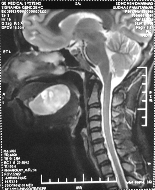
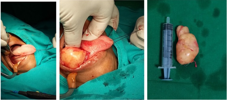
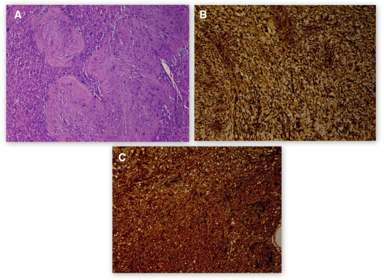
Ancient schwannoma is an uncommon schwannoma variant which is a benign tumour of Schwann cell origin. It is relatively a rare tumour (25%) in the head and neck region. It is encapsulated and well demarcated from the surrounding tissues. A 35 years old patient in this case presented with painless swelling of the tongue. Trans-oral excision of the intramural lesion was performed. Histopathological and immuno-histochemistry confirmed the diagnosis of schwannoma. This case is of interest on account of the complexity of its diagnosis and the atypical site, that is, intra-mural, of appearance of a schwannoma.

Keywords: Ancient schwannoma; Antoni A, Antoni B bodies; Intramural intra-lingual mass; Tongue swelling.


Conflict of interest statement

Conflict of interestAll authors declare that they have no conflict of interest.

Figures

Fig. 1
Fig. 1
A 4 × 4 cm solitary, spherical, smooth swelling situated over the dorsum of the tongue
Fig. 2
Fig. 2
T1W STIR image showing a hyperintense lesion with internal cystic changes measuring 3.1 cm cranio-caudal (CC) × 5.0 cm antero-posterior (AP) × 3.2 cm medial-lateral (ML)
Fig. 3
Fig. 3
Showing trans oral excision of lesion was done via a 5 cm incision over the right lateral border. Yellowish encapsulated mass excised en-bloc from within the musculature
Fig. 4
Fig. 4
a Histopathology showing a well encapsulated lesion comprising of spindle shaped cells arranged in fascicles showing spindle to ovoid nuclei and few bizarre nuclei with few hypo cellular areas and sparse mitotic activity and Antoni Type A and Type B cells with Verocay bodies with hyalinised vessels. b Immuno-histochemistry showing S-100 positive. c Vimentin markers positive
Fig. 5
Fig. 5
On post—op 2 months follow-up

References

    1. Enzinger FM, Weiss SW. Soft tissue tumors. 3. St Louis: Mosby-Year Book Inc.; 1995. pp. 821–888.
    1. Karaca CT, Habesoglu TE, Naiboglu B, Habesoglu M, Oysu C, Egeli E, Tosun I. Schwannoma of the tongue in a child. Am J Otolaryngol. 2009;31:46–48. doi: 10.1016/j.amjoto.2008.09.010. - DOI - PubMed
    1. Jones JA, McWilliam LJ. Intraoral neurilemmoma (schwannoma): an unusual palatal swelling. Oral Surg Oral Med Oral Pathol. 1987;63(3):351–353. doi: 10.1016/0030-4220(87)90203-9. - DOI - PubMed
    1. Wayne Lollar K, Pollak N, Daniel Liess B, Miick R, Zitsch R., III Schwannoma of the hard palate. Am J Otolaryngol. 2010;31:139–140. doi: 10.1016/j.amjoto.2008.11.009. - DOI - PubMed
    1. Moreno-García C, Pons-García MA, González-García R, Monje-Gil F. Schwannoma of tongue. J Maxillofac Oral Surg. 2014;13(2):217–221. doi: 10.1007/s12663-010-0101-0. - DOI - PMC - PubMed

LinkOut - more resources